Tissue and Cell Imaging by IR Lasers
What is spatial biology?
In living organisms, cells interact and assemble in three-dimensional tissues. The spatial orientation and position of a cell is critical for its function, development and interaction with the surrounding environment. Spatial biology focuses on visualizing cells and molecules to investigate how these spatial distributions influence underlying biological processes and mechanisms. Therefore, spatial biology promises to revolutionize health and disease research.
Key methods in spatial biology include advanced imaging technologies, such as infrared (IR) imaging and matrix-assisted laser desorption/ionization (MALDI) imaging that enable the mapping of molecular distributions at the low-micrometer-scale without the requirement of labelling or tagging. Yet, limitations in speed and throughput hindered the analysis of large tissue sections and/or substantial sample numbers.
Now, the diversity and complexity of biological tissues, i.e. cell type, microenvironment, and structural features, can be captured at ultra-fast speed and with high precision. IR laser imaging significantly outperforms the measurement speeds of conventional FT-IR imaging, simultaneously providing breathtaking images.
IR Laser Imaging goes beyond limitations in tissue analysis
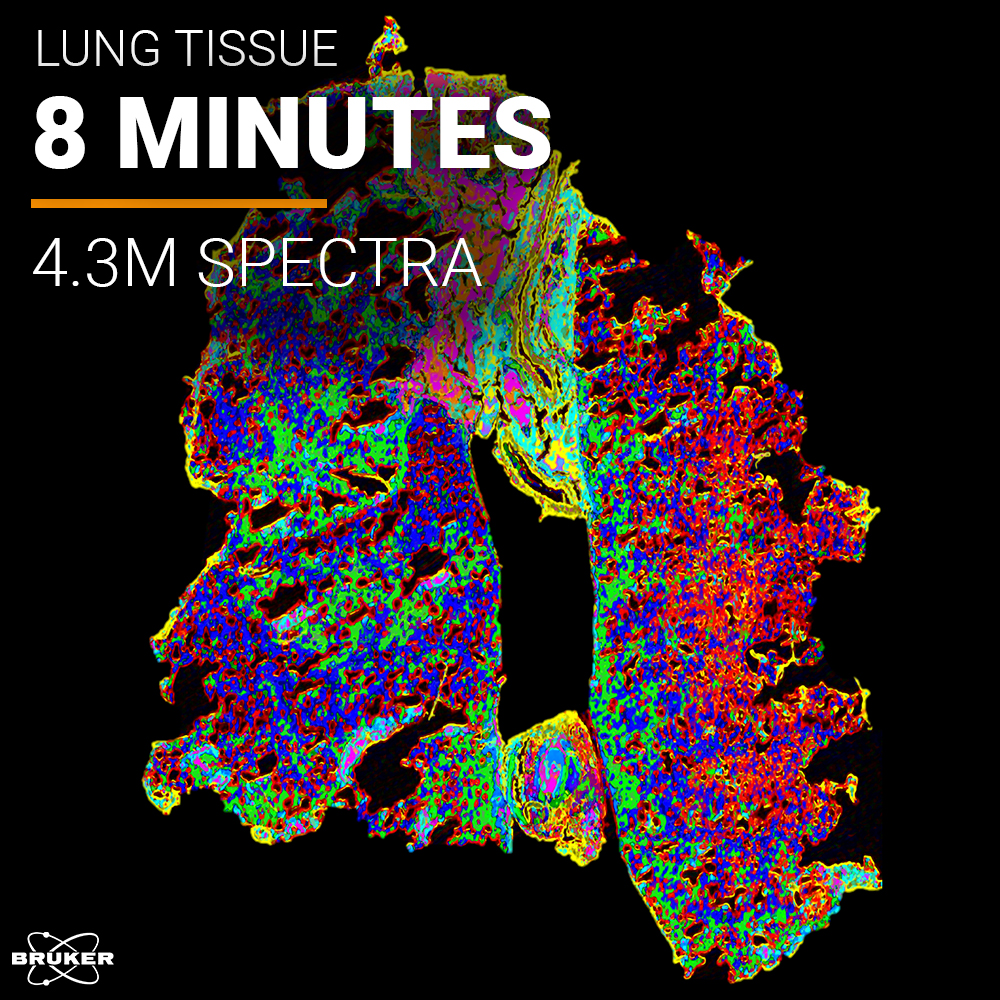
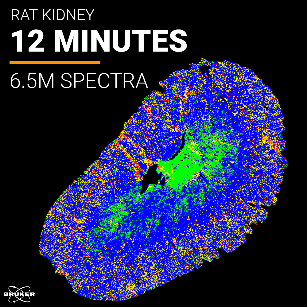
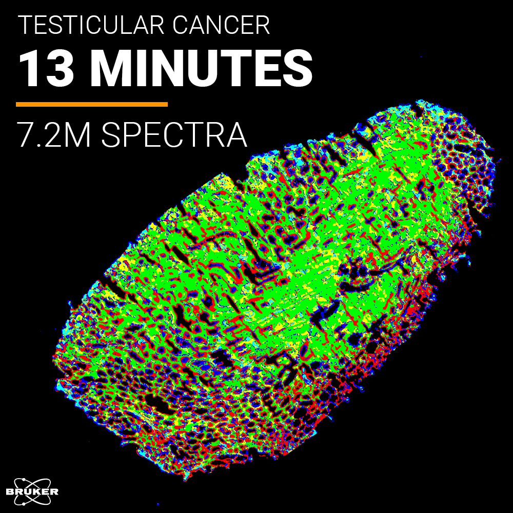
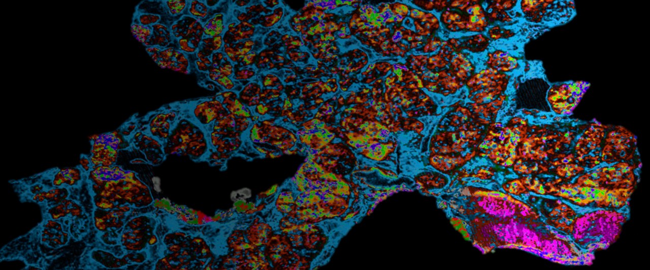
By using IR lasers, tissue imaging has become faster than ever. With acquisition rates of 9000 pixels per second at a pixel resolution of 5 µm, Bruker's ILIM allows the imaging of several square centimeters of tissue within minutes. By implementing our patented coherence reduction technique, it is finally possible to capture theses images without artifacts – something no other IR instrument can achieve. Adding to this, our unique machine learning-driven algorithms generate spatial segmentation maps based on chemical fingerprints without the need for prior molecular or histological input. The result is an unprecedented analytical tool for tissues that opens new possibilities and applications, such as IR Guided MALDI Imaging.
Introducing IR Guided MALDI Imaging
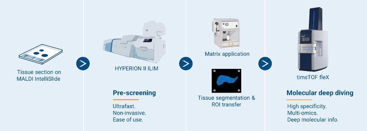
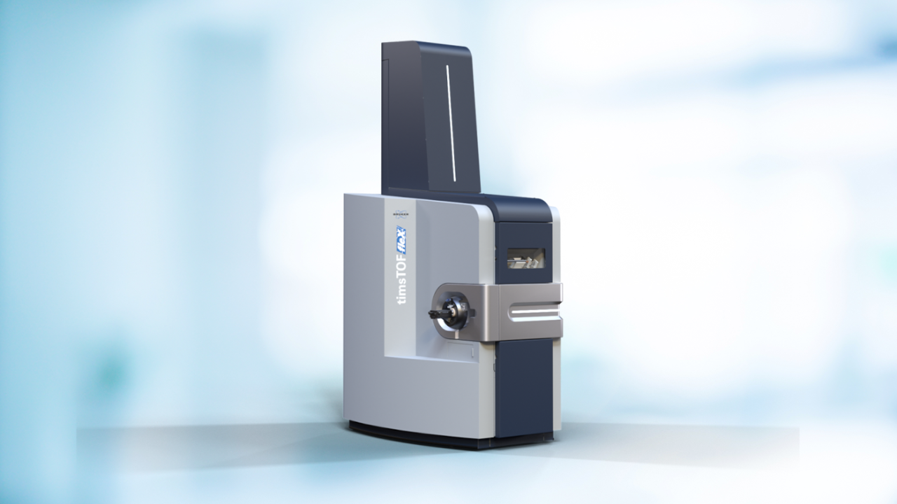
Breakthrough IR Laser Imaging performance is combined with best-in-class MALDI Imaging. The new seamless end-to-end workflow for IR Guided MALDI Imaging features the HYPERION II ILIM and the timsTOF fleX – letting you power through spatial biology studies on an unprecedent scale.
No modifications or additional steps regarding sample preparation are required and both modalities are conducted on the same tissue section. On a minute time scale, get comprehensive tissue segmentation maps by IR Laser Imaging, transfer the spatial information and perform guided MALDI Imaging of exclusive tissue features to reduce acquisition times – thereby significantly increase throughput and work with sample numbers and sizes that were previously impossible to tackle.
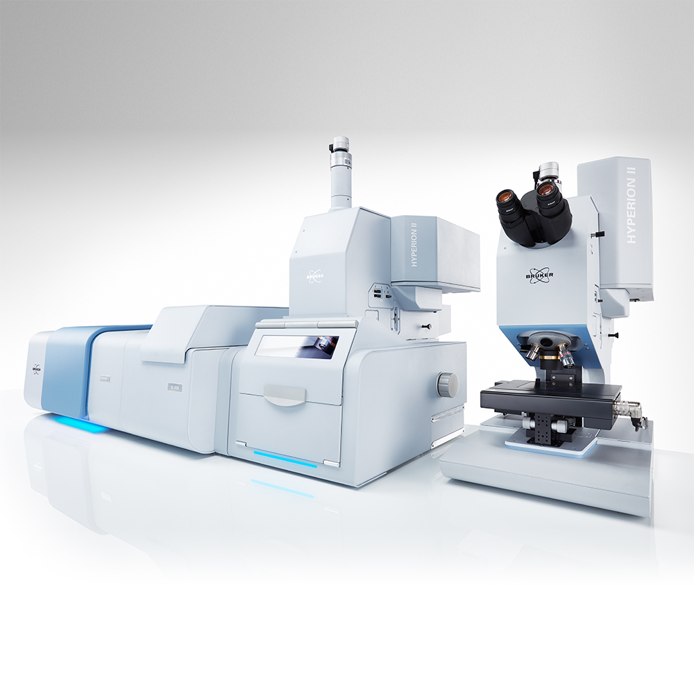
Meet the ultimate solution to tissue and cell imaging
Our pioneering QCL technology delivers exceptional IR laser imaging performance that is up to 180 times faster than traditional FT-IR Imaging. Innovative hardware design for coherence reduction to acquire artifact-free IR Imaging data with high fidelity unlike any other IR instrument - all while still being augmented by FT-IR.
Dual ion source design brings unparalleled versatility to enable ESI/MALDI with zero compromises. More molecular information behind every pixel with SpatialOMx. TIMS to solve the highest molecular complexity. Smartbeam 3D and microGRID for unmatched spatial resolution.