Vutara VXL
Vutara VXL
The Bruker Vutara VXL super-resolution microscope enables researchers to enter the world of super-resolution imaging with ease. It incorporates Bruker’s industry-leading single-molecule localization microscopy (SMLM) technology in a streamlined system with a compact footprint. The Vutara VXL system allows you to perform research on DNA, RNA, and proteins, from macromolecular complexes and super-structures to chromatin structure and chromosomal substructures, as well as studying functional relationships in genomes and various subcellular organelles. This novel system also supports advanced spatial biology research in extracellular matrix structures, extracellular vesicles (EV), virology, neuroscience, and live-cell imaging. When combined with Bruker's unique microscope fluidics unit, Vutara VXL enables multiplexed imaging for targeted, sub-micrometer multiomics in genomics, transcriptomics, and proteomics research.
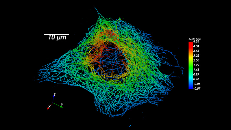
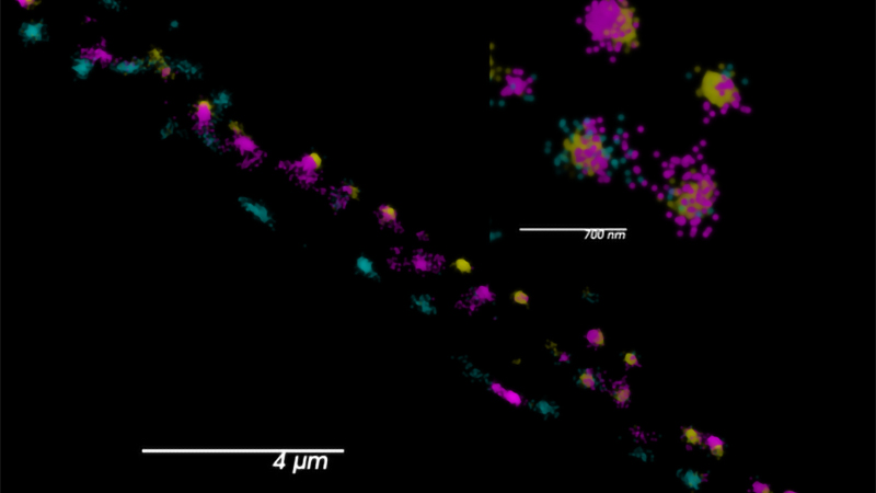
Every Acquisition Is 3D
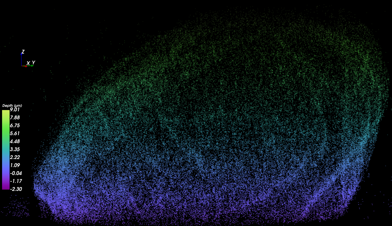
The Vutara VXL is equipped with proprietary bi-plane technology that enables you to gather 3D data with every acquisition. This technology allows you to easily perform a Z-stack series for thicker specimens and automatically localize and reconstruct the entire volume. With Vutara VXL, you can obtain comprehensive 3D data of your samples with ease.
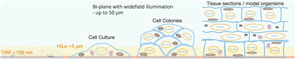
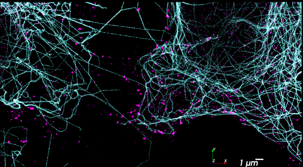
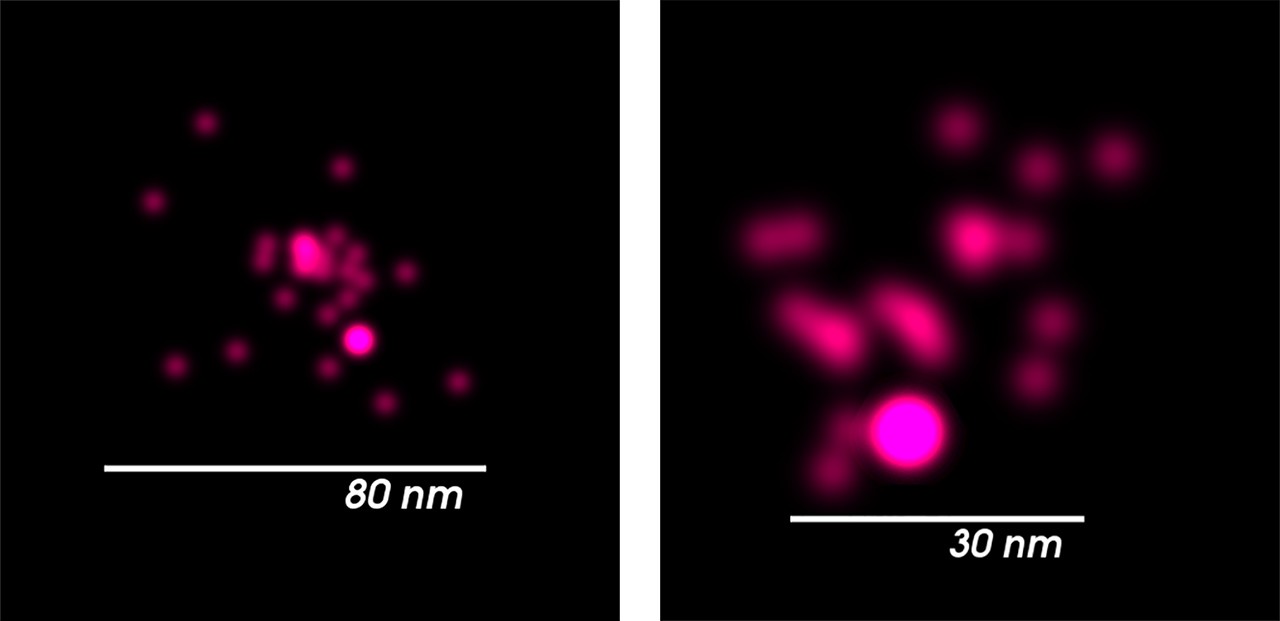
Single-Molecule Imaging Beyond the Cover Slip
The Vutara VXL is capable of imaging far from the surface of the coverslip, about 50 µm deep, to accommodate a wide range of sample types. Thanks to the proprietary biplane technology, the Vutara VXL with the SRX software can perform single-molecule localization microscopy on more sample types than any other commercial single-molecule localization microscope available. Making cultured cells, cell colonies, tissue sections, and entire model organisms accessible for your single-molecule localization experiments.
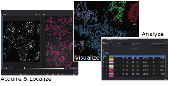
Turn Localizations Into Information
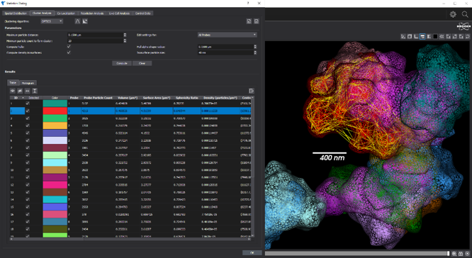
Vutara’s Quantitative Localization Microscopy suite enhances your productivity and allows you to turn localizations into meaningful results. Vutara's SRX workflow-driven software guides users through the setup, calibration, imaging, processing, and analysis of the super-resolution single-molecule localization experiment. SRX boosts the value of the system by providing dedicated software that is designed specifically for SMLM. The SRX software combines real-time localization processing with advanced visualization and sophisticated quantitative analysis tools to let researchers quickly create publication quality videos, images, and measurements.
Specifications
| Super-resolution localization microscopy (SMLM) |
|
| Widefield microscopy |
|
| Excitation lasers (nominal laser power at diode) |
|
| Flat illumination |
|
| Multi-color acquisition |
|
| Multi-plane imaging |
|
| Camera |
|
| Objective |
|
| Field of View (FOV) |
|
| 3D SMLM method |
|
| SMLM resolution |
|
| Imaging depth |
|
| xy-stage |
|
| z-focus |
|
| Drift correction |
|
| Environmental control |
|
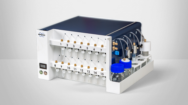
| Microfluidics unit for sequential labeling |
|
| Compact table-top design |
34 cm x 40 cm x 34 cm, (1.2 ft x 1.3 ft x 1.2 ft) 35 kg (78 lbs)
33 cm x 53 cm x 32 cm (1.0 ft x 1.7 ft x 1.0 ft) 30 kg (65 lbs)
53 cm x 35 cm x 14 cm (1.7 ft x 1.1 ft x 0.4 ft) 8 kg (17 lbs) |
| Operating in a typical laboratory environment |
|
| Vibration insulation included |
|
| Workflow-defined software for easy data acquisition |
STORM / dSTORM PALM PAINT Other blinking-based modalities
Chromatin tracing smFISH
Tracking of probe cycles and positions Support for an unlimited number of probes
|
| SMLM processing |
|
| Particle tracking |
|
| Fluidics Manager |
|
Drift correction |
|
| Statistical analysis tools |
Ripley’s K functions Pair correlation Nearest neighbors
DBSCAN OPTICS Delaunay Particle counts Volume and density calculations Determination of radius of gyration and sphericity Principal component analysis (PCA) Calculation of centroid
Pearson’s correlation coefficient Mander’s overlap coefficient Intensity correlation quotient Cross pair correlation Joint histogram STORM-RLA
Fourier ring correlation Labeling density resolution analysis Local resolution correlation mapping Nearest neighbors’ analysis |
| Chromatin tracing (optional) |
|
| Integrated visualization |
|
| Open data formats |
|
| Network attached storage (NAS) unit (optional) |
|
A Complete Solution, From Acquisition to Analysis
With SRX software and its Quantitative Localization Microscopy analysis suite, Vutara VXL can provide visual and quantitative information from biological samples. By localizing individual molecules, Vutara can generate stunning 3D images while simultaneously providing in-depth quantitative analysis tools.
Recent Webinars
With more than 15 years of experience in designing microscopes in academia and in industry, I am proud to contribute to the development of Vutara's super-resolution microscopes. With biplane 3D detection and fast sCMOS imaging, Vutara has the most advanced super-resolution microscope on the market.

After scanning the market for super-resolution microscopes and personally visiting and testing most commercially available systems with our own samples, I can say that I am most impressed with Vutara's SR-350. In particular, the user friendliness of their imaging software as well as the 3D capability in super-resolution mode impressed me.
Vutara has an excellent support team and staff as a whole making our transition to super-resolution a well-supported experience. If you're looking to advance your research by super resolution microscopy, I can confidently say that Vutara's systems are an excellent choice.

The volume of data you can acquire on a super-resolution system is incredible when comparing electron microscopy especially considering the sample preparation.

Having published with a Vutara super-resolution system, I can confidently say that they offer one of the most advanced super-resolution microscopes available. Their attention to detail and ongoing close collaboration makes them one of my preferred microscopy vendors.