
Interferometric Nanoparticle Tracking Analysis (iNTA)
Label-Free
Nanoparticle Tracking
What Is Nanoparticle Tracking Analysis (NTA)? What is interferometric NTA?
Nanoparticle Tracking Analysis (NTA) is a single-particle measurement technique used to determine nanoparticle size and concentration in liquid samples. Individual particles are tracked as they undergo Brownian motion, and their movement is used to calculate particle size.
Interferometric Nanoparticle Tracking Analysis (iNTA) extends this approach by using interferometric detection. In addition to particle size and concentration, iNTA measures optical contrast for individual particles, providing an additional parameter that can help differentiate particle populations in heterogeneous samples.
How Does Interferometric Nanoparticle Tracking Analysis (iNTA) Work?
iNTA tracks individual nanoparticles in liquid while measuring their interferometric optical contrast. Particle motion is used to determine particle size, while the optical contrast provides an additional particle-specific measurement parameter. Combining these measurements makes it possible to characterize heterogeneous nanoparticle populations with more information than size alone.
NTA vs. iNTA: What Is the Difference?
| NTA (darkfield) | iNTA (interferometry) | |
| Size Resolution | good | very good |
| Concentration | single population | multi population |
| Particle Properties | size | size + refractive index |
| Plate Reader | no | yes |
| Well Plate Format | - | 18-well plate |
| Sample Volume | 250–1,000 µL | 50–100 µL |
| Sample Conservation | low | high |
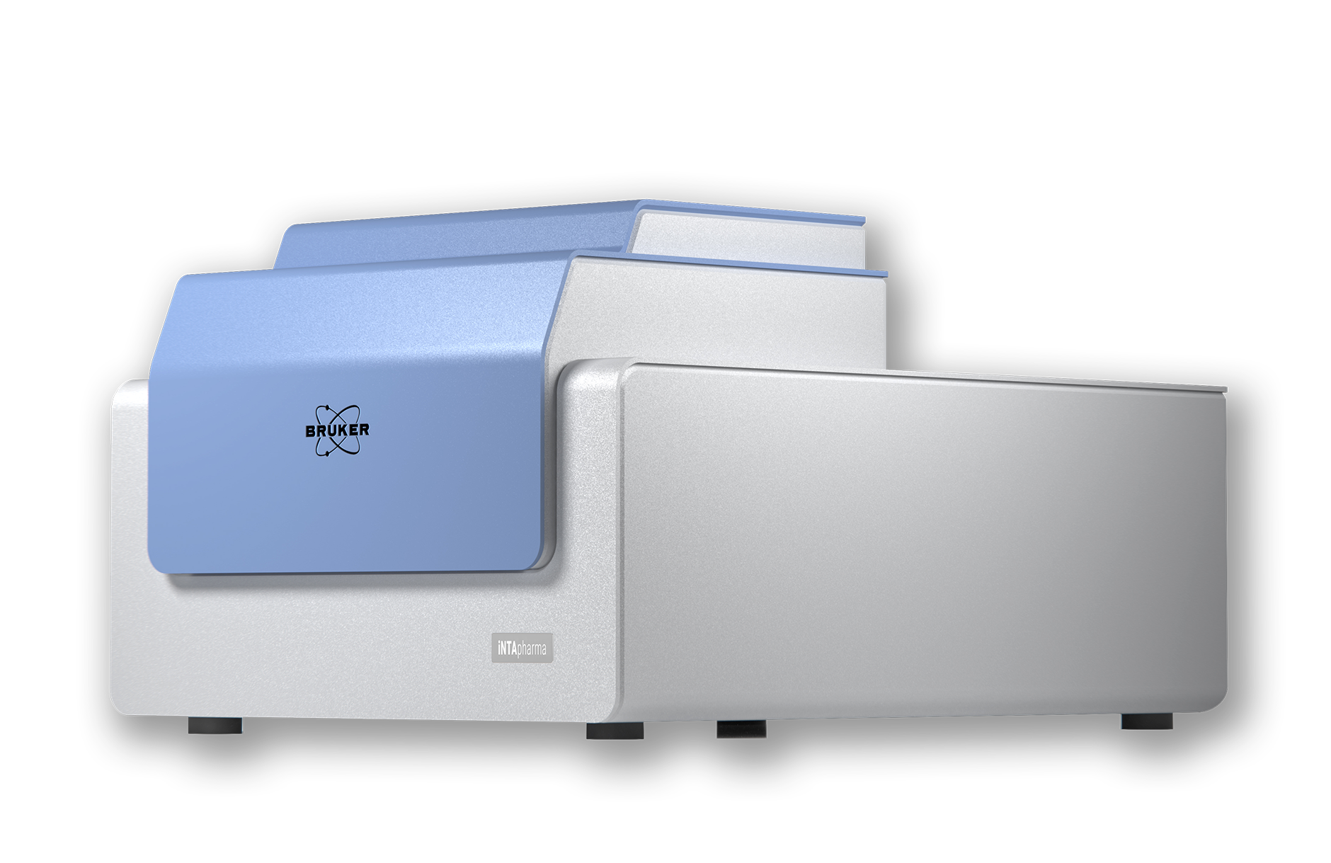
Meet iNTApharma: interferometric Nanoparticle Tracking Analyzer
iNTApharma is Bruker’s automated interferometric nanoparticle tracking analyzer for label-free, single-particle characterization. It measures particle size, concentration, and optical contrast using low sample volumes and an 18-well consumable workflow.
By combining particle size with interferometric optical contrast, iNTApharma provides a two-dimensional view of heterogeneous particle populations. It supports the characterization of extracellular vesicles, exosomes, lipid nanoparticles, and viral vectors, including populations that may overlap in size.
The 18‑well plate consumable simplifies your experiments.

Fill consumable in biosafety cabinet and seal it.

Place consumable on instrument and start analysis.

Assess Critical Quality Attributes

Easy disposal of consumable after autoclaving it for 30 min.

Exosome Analysis by iNTA
What Are Exosomes?
Exosomes are a subtype of extracellular vesicles (EVs), a heterogeneous class of membrane-bound particles released by cells into the extracellular environment. Typically ranging from 30–150 nm in diameter, exosomes originate from the endosomal pathway and are secreted through the fusion of multivesicular bodies with the plasma membrane. While EVs also include microvesicles and apoptotic bodies, exosomes have emerged as a major focus of research due to their role in cell-to-cell communication and their potential as disease biomarkers.
Why Analyze Exosomes with iNTA
The isolation of exosomes often results in complex samples that may also contain lipoproteins, protein aggregates, and other nanoparticle populations overlapping in size. Conventional size-based characterization alone can therefore be insufficient to fully understand sample composition.
By combining particle tracking with interferometric optical contrast, iNTA adds an additional analytical dimension for exosome characterization:
- Label-Free Population Analysis: Differentiate exosome-related particle populations from co-isolated nanoparticles using size and interferometric optical contrast.
- Exceptional Resolution: Resolve exosome size distributions with a level of detail comparable to electron microscopy, while maintaining the advantages of solution-based measurements.
- Saves you time and sample: Perform repeated measurements using low sample volumes, automated plate-based handling, and an integrated 18-well workflow.

Go Beyond Counting and Sizing
iNTApharma extends traditional exosome analysis beyond particle concentration and size determination. By visualizing individual particles according to both size and optical contrast, researchers can investigate sample heterogeneity and identify which particle populations contribute to the overall measurement.
Rather than relying solely on one-dimensional size distributions, iNTA provides multidimensional insight into complex exosome preparations.
Application Note: Label-Free Discrimination of Exosomes

This application note highlights how interferometric Nanoparticle Tracking Analysis (iNTA) achieves label-free discrimination of exosomes from co-isolates like lipoproteins in complex biological fluids. It is a vital read for researchers seeking to bypass the limitations of traditional size-based separation and fluorescence labeling.
Viral Vector Analysis by iNTA
What Are Viral Vectors?
Viral vectors are engineered viruses used to deliver genetic material into cells and are a foundational technology in gene therapy, cell therapy, and vaccine development. Common vector platforms include adenoviral vectors (AdV), adeno-associated viruses (AAV), and lentiviral vectors.
The successful development and manufacturing of viral vectors depend on accurate characterization of critical quality attributes (CQAs), including particle size, concentration, purity, and the presence of unwanted particle populations. Reliable analytical methods are therefore essential for ensuring product quality and process consistency.
Why Analyze Viral Vectors with iNTA?
Viral vector samples can contain heterogeneous particle populations, aggregates, contaminants, and other particles with overlapping size distributions. Size-based characterization alone may therefore provide an incomplete picture of sample composition and quality.
By combining particle tracking with interferometric optical contrast, iNTA adds an additional analytical dimension for viral vector characterization:
- Particle-by-Particle Analysis: Measure individual particles by size and interferometric optical contrast to better characterize heterogeneous viral vector samples.
- Population Differentiation: Identify particle populations that may overlap in size but differ in optical properties, supporting a more detailed assessment of sample composition.
- Low-Volume, Automated Measurements: Analyze samples using low volumes and an automated 18-well plate workflow, supporting research, process development, and quality assessment.

Go Beyond Size and Particle Counts
Traditional particle analysis methods often rely solely on size distributions, making it difficult to identify subpopulations with similar sizes but different physical characteristics.
iNTA visualizes every tracked particle according to both size and optical contrast, creating a multidimensional representation of the sample. This allows researchers to distinguish intact viral vectors from contaminants or other particle populations and evaluate sample purity on a particle-by-particle basis.
Rather than relying on one-dimensional size information alone, iNTA provides a more complete picture of viral vector quality and sample complexity.
Application Note: Quality Assessment of Adenoviral Vectors

This application note details how interferometric Nanoparticle Tracking Analysis (iNTA) provides high-resolution, label-free quality control for adenoviral vectors (AdV). It is essential for researchers needing to distinguish intact viruses from contaminants or empty capsids, a task challenging for conventional methods like DLS or NTA.
Recent Publications on iNTA

Employing high‑resolution interferometric Nanoparticle Tracking Analysis to investigate how freezing‑induced accelerated ageing enhances the homogeneity, stability, and reproducibility of red blood cell‑derived extracellular vesicles for biomaterial applications.
Learn more here:
Paolini, L., Romano, M., Mangolini, V., Tassoni, S., Jiang, S., Mazzoldi, E. L., Musicò, A., Zendrini, A., Kashkanova, A., Sandoghdar, V., Berardi, A. C., Giliani, S. C., Bergese, P., Radeghieri, A.
Biomaterials Science, 14, 122–139, 2026

Employing interferometric Nanoparticle Tracking Analysis (iNTA) to enable quantitative, calibration‑free measurements of nanoparticle concentrations in complex and polydisperse liquid mixtures.
Learn more here:
Measuring Concentration of Nanoparticles in Polydisperse Mixtures Using iNTA
Kashkanova, A. D., Albrecht, D., Küppers, M., Blessing, M., Sandoghdar, V.
ACS Nano, published online July 9, 2024

Applying interferometric Nanoparticle Tracking Analysis for label‑free differentiation and accurate quantification of extracellular vesicles in complex biological samples containing large lipoproteins.
Learn more here:
Label‑free discrimination of extracellular vesicles from large lipoproteins
Kashkanova, A. D., Blessing, M., Reischke, M., Baur, J.-O., Baur, A. S., Sandoghdar, V., Van Deun, J.
Journal of Extracellular Vesicles, 12(8), 2023

Employing interferometric Nanoparticle Tracking Analysis (iNTA) to enable high‑precision, non‑invasive measurement of size and refractive index distributions in weakly scattering nanoparticles within complex and polydisperse liquid samples.
Learn more here:
Precision size and refractive index analysis of weakly scattering nanoparticles in polydispersions
Kashkanova, A. D., Blessing, M., Gemeinhardt, A., Soulat, D., Sandoghdar, V.
Nature Methods, 19(5), 586–593, 2022
Get in touch with our iNTA experts
Directly talk to our application experts and learn how iNTA can support your work.