Biomarkers for Huntington's Disease
What is, in your opinion, the most significant achievement in Huntington’s disease research in the past 3 years?
Huntington’s disease (HD) is an inherited neurodegenerative disorder characterized by involuntary abnormal movements, as well as cognitive and psychiatric symptoms associated with early atrophy of the striatum and cerebral cortex. The mutation causing HD consists of an abnormal expansion of a CAG repeat in the gene coding the protein huntingtin (htt). This leads to an expansion of a poly-glutamine tract in the N-terminal part of the protein that is believed to be toxic to brain cells, leading to neurodegeneration. Despite tremendous efforts made, there is currently no therapy to cure the disease or to slow its progression.
Nonetheless, new molecular biology techniques aiming at reducing mutant htt levels have emerged in the past years. Selective gene silencing has shown therapeutic efficacy in both cellular and mouse models of HD. This opens very promising ways to stop or to slow down the course of the disease in humans.
What do believe the next great step forward in preclinical Huntington’s disease research will be?
As mentioned, gene-silencing or gene-suppressive strategies such as ASO (antisense oligonucleotides), siRNA (small interfering RNA) or CRISPR/Cas9 are very promising to directly modify the course of the disease. Several seminal works have demonstrated therapeutic efficacy of such approaches and some of them have already been transfer to clinical trials. To date, the atrophy of the striatum as measured by MRI is currently the best biomarker of disease progression in HD gene carriers. However, anatomical measurements are too limiting to capture the extent of different biological processes involved in HD. Thus, there is an urgent need to find more predictive, functional and earlier biomarkers. Preclinical research involving animal models of HD associated with high performance neuroimaging modalities are absolutely crucial for biomarker identification and characterization. Moreover, in the perspective of future treatments, such biomarkers would be extremely useful to evaluate drug engagement and biological efficacy.
Could you refer a good review paper of preclinical MRI research on Huntington’s disease?
The reader could refer to the review [1]
- Novel Imaging Biomarkers for Huntington’s Disease and Other Hereditary Choreas. Fazio P., Paucar M., Svenningsson P., Varrone A., Current Neurology and Neuroscience Reports (2018) 18: 85,
https://www.ncbi.nlm.nih.gov/pmc/articles/PMC6182636/
doi: 10.1007/s11910-018-0890-y
or to the book chapter [2]
- In Vivo Multidimensional Brain Imaging in Huntington's Disease Animal Models. Flament J., Hantraye P., Valette J., Methods Mol Biol (2018) 1780:285-301.
https://pubmed.ncbi.nlm.nih.gov/29856025/
doi: 10.1007/978-1-4939-7825-0_15
Can you explain the role that glutamate plays in Huntington’s disease and how this is being currently investigated?
Glutamate is the major excitatory transmitter in the central nervous system and is involved in several aspects of normal brain functions including cognition, memory and learning. In normal conditions, most of glutamate is located in cells, neurotransmission being governed by a few micromolar of extracellular glutamate. Glutamate can become toxic if extracellular concentration is too high. Thus, mechanisms to maintain low extracellular concentration are crucial for brain functions. In addition, glutamate is central to several metabolic pathways related to energy metabolism and oxidative stress so its concentration has to be precisely regulated and a small defect can be very deleterious for the brain.
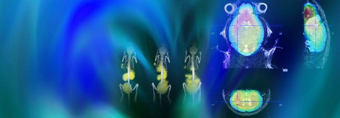
Recently, CEST (Chemical Exchange Saturation Transfer) has been proposed to address the sensitivity limitation of in vivo 1H Magnetic Resonance Spectroscopy by using indirect detection of dilute molecules through their exchange of labile protons with the bulk water. The potential of CEST imaging to map glutamate level (gluCEST) has already been demonstrated in both rodent and human brains at high magnetic fields (≥ 7T). In the context of HD, gluCEST has already permitted to highlight significant decreases of glutamate levels in several brain regions in a mouse model of HD [3]. More interestingly, the most affected structure was the corpus callosum, suggesting that this structure was very early affected in this mouse model. In addition, a longitudinal and multimodal MRI approach, combining Diffusion Tensor Imaging (DTI), Magnetization Transfer (MT) and gluCEST has pointed out the potential role of white matter in the brain dysfunction that characterize HD and highlighted the pertinence of gluCEST as biomarkers in HD [4].
How is an increased understanding of brain alterations via MR imaging able to translate into patient treatment?
Pushing the limits of preclinical imaging methods in control animals and in animal models of neurodegenerative disorders is crucial to better understand normal brain physiology and to elucidate biological processes in pathological conditions. In addition, it provides tools to accurately evaluate therapeutic efficacy of future treatments. Moreover, with the increasing availability of high field clinical scanners, most of MRI modalities initially developed on preclinical scanners will be transferable for clinical evaluation and to assess disease progression in a clinical context of HD. This might help identifying more relevant and early biomarkers in premanifest HD patients and could provide relevant information about disease pathogenesis.
References
[1] Novel Imaging Biomarkers for Huntington’s Disease and Other Hereditary Choreas.
Fazio P., Paucar M., Svenningsson P., Varrone A., Current Neurology and Neuroscience Reports (2018) 18: 85.
https://www.ncbi.nlm.nih.gov/pmc/articles/PMC6182636/
doi: 10.1007/s11910-018-0890-y
[2] In Vivo Multidimensional Brain Imaging in Huntington's Disease Animal Models.
Flament J., Hantraye P., Valette J., Methods Mol Biol (2018) 1780:285-301.
https://pubmed.ncbi.nlm.nih.gov/29856025/
doi: 10.1007/978-1-4939-7825-0_15
[3] In vivo imaging of brain glutamate defects in a knock-in mouse model of Huntington's disease.
Pépin J., Francelle L., Carrillo-de Sauvage M-A., de Longprez L., Gipchtein P., Cambon K., Valette J., Brouillet E., Flament J., NeuroImage (2016) 139:53-64.
https://pubmed.ncbi.nlm.nih.gov/27318215/
doi: 10.1016/j.neuroimage.2016.06.023
[4] Longitudinal multimodal MRI characterization of a knock-in mouse model of Huntington’s disease reveals early grey and white matter alterations.
Pérot J-B., Célestine M., Palombo M., Dhenain M., Humbert S., Brouillet E., Flament J., Human Molecular Genetics (2022) 31:3581–3596.
https://pubmed.ncbi.nlm.nih.gov/35147158/
doi: 10.1093/hmg/ddac036
Despite tremendous efforts made, there is currently no therapy to cure the disease or to slow its progression.
Preclinical research involving animal models of Huntington’s disease associated with high performance neuroimaging modalities are absolutely crucial for biomarker identification and characterization.
Dr. Julien Flament
Dr. J. Flament is researcher at the Molecular Imaging Research Center (MIRCen) - CEA located in Fontenay-aux-Roses, France. His personal research is focused on development of metabolic imaging using CEST technic at very high magnetic field to better understand brain energy metabolism in normal and pathological conditions, especially in the context of neurodegenerative diseases.