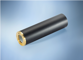
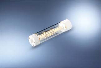
Micro5
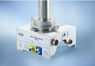
Microscopy images with resolutions < 10 x 10 x 10 μm3 samples in a Bruker standard bore magnet. But the probe can also be used in wide bore and super wide bore systems. Combined with it‘s versatile exchangeable rf coils it can be adapted to perfectly match your application, supporting sample diameters from 0.5 -10 mm.
The Micro5 probe can also be used as a 3-axis diffusion probe or combined to increase the gradient strength in one direction. It is particularly useful to investigate anisotropic diffusion.
Features
- Standard Bore Probe, also compatible to WB and SWB magnets
- Gradient strength of up to 3 T/m, water cooled
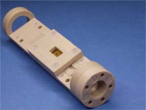
- Exchangeable rf coils
- Micro5 rf coils (compatible to Diff50 except 10 mm coils)
2 and 4mm Solenoid
- 5-10 mm Volume Coils (Sample transfer via SB-BST)
- 500 μm MicroCoils
- Double channel 5-8 mm coils and single channel 5-10 mm coils
- Variable Temperature Control (compatible to BSVT and BCU)
- Compatible to SB or WB Shim System
- Compatible to TopShim
- Probe body compatible to Diff50 MR Diffusion Probe
Specifications
| Micro5 | |
| Bore | SB / WB / SWB |
| 1H frequency range | 300 – 950 MHz |
| rf channels | Two independent |
| Requirements | MR Microscopy Accessory |
| Gradient | Micro5 |
| Directions | xyz |
| Gradient strength per direction | 3 T/m |
| ID/OD | 19/40 mm |
| Rise Time (5-95%), 0-60A,120V | < 80 µs |
| Cooling | Water |
| Maximum current tested | 60 A |
| Removable | Yes |
| Exchangable rf coil | |
| Detection | single tuned, double tuned |
| Typical Nuclei | 1H, 2H, 19F, 13C, 31P, 7Li, 19F, … |
| Standard sample temperature range (5 mm) | -40°C.. +80°C |
| Sample diameter | 0.5 – 10 mm |
Applications
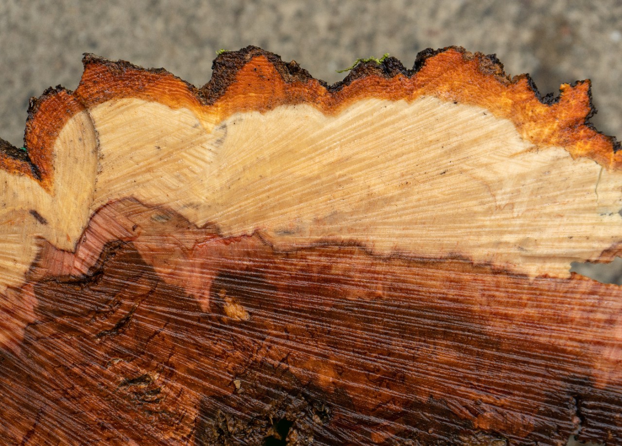
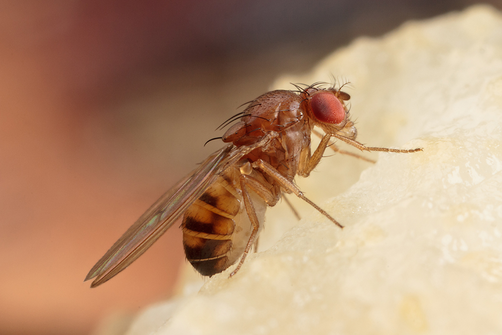
The Micro5 probe is typically used to study mineral or geologic samples, polymers, tablet dissolution kinetics, plants and seeds, bone or tissue biopsy samples, and small insects.
Accessories
Rf coils are exchangeable, the gradient coil must be unmounted first to allow the change of the rf coil.
- 1 mm MicroCoil (coil diameter 1 mm, max. sample size 500 μm). To replace the sample the probe must be removed from the magnet as with the solenoid coils, unless e.g. a Flow Cell is used. Different Type of sample containers are supported
- 2-4 mm Solenoid coil. The samples must be placed horizontally in this type of rf coils. This requires to remove the probe from the magnet, remove the gradient coil and place to sample.
- 5 or 10 mm single tuned saddle coil or a 5 or 8 mm double tuned coil. In this case the sample can be changed using the Bruker BST sample transfer system, the probe remains inside the magnet.
- Diff Software Application
- Dynamic Center for data processing
Labscape
Service & Life Cycle Support for Magnetic Resonance and Preclinical Imaging
Bruker’s commitment to provide customers with unparalleled help throughout the buying cycle, from initial inquiry to evaluation, installation, and the lifetime of the instrument is now characterized by the LabScape service concept.
LabScape Maintenance Agreements, On-Site On-Demand and Enhance Your Lab are designed to offer a new approach to maintenance and service for the modern laboratory


MR microscopy & MR diffusion applications team: micdiff@bruker.com