
Mouse Imaging
The mouse and humans share a common ancestor, thus it is of no surprise, that their genes and genomes are very similar. Due to this similarity, the mouse is used as a model organism to study human biology. With the help of the mouse diseases, such as cancer, cardiovascular diseases or brain disorders, can be researched.
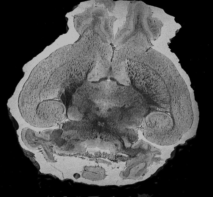
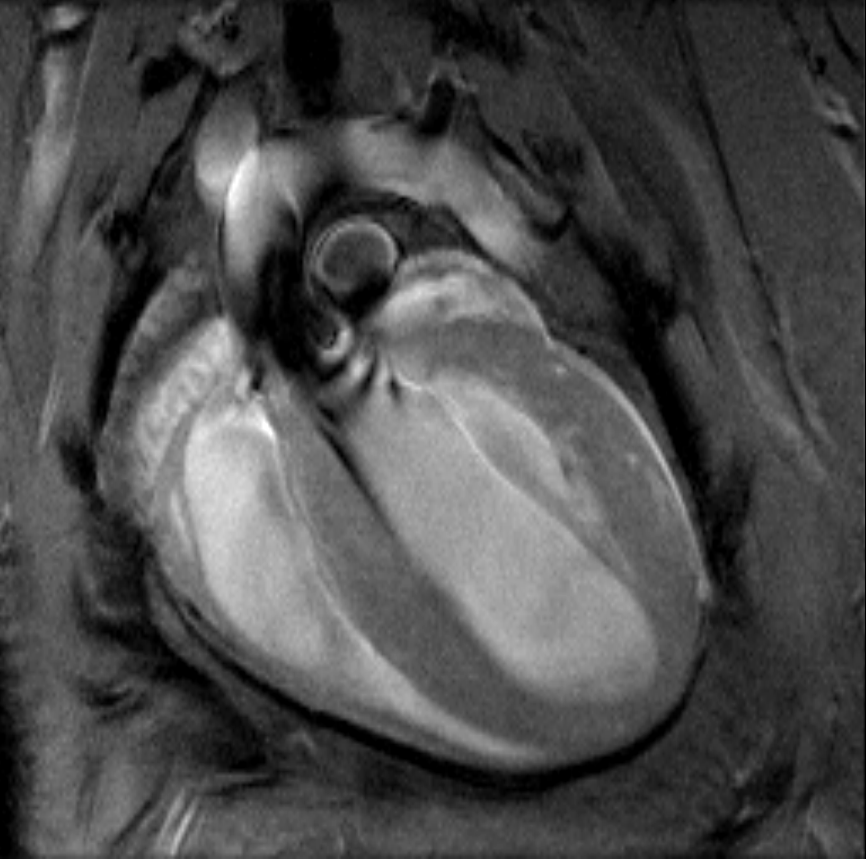
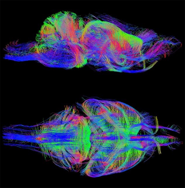
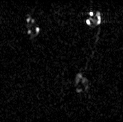
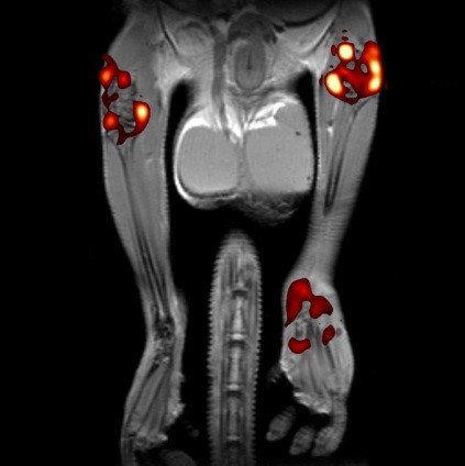
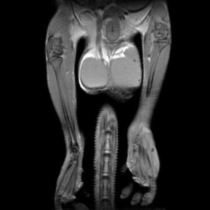
MR microscopy enables to acquire in-vivo images of the neuronal pathways in the brain (Figure 2& Figure 3) and even record videos of a beating heart (Figure 3), where each single image of the video sequence can be triggered by EKG, for better image resolution. Additional marker substances enable highlighting area such as the inflammations in Figure 4.
References:
Bruker Application note: Cardiovascular Magnetic Resonance in Mice, Ulrich Flögel, Experimental Cardiovascular Imaging Heinrich Heine University, Düsseldorf, Germany floegel@uni-duesseldorf.de,
https://www.google.com/url?sa=t&rct=j&q=&esrc=s&source=web&cd=&cad=rja&uact=8&ved=2ahUKEwiu0OX2mNbpAhXnUBUIHQarClUQFjABegQIAxAB&url=https%3A%2F%2Fwww.bruker.com%2Ffileadmin%2Fuser_upload%2F8-PDF-Docs%2FPreclinicalImaging%2FT177101_App_Note_Cardiovascular_Magnetic_Resonance_in_Mice.pdf&usg=AOvVaw3wGL-QsA312ORCKC9Ho5qp (better link needed)
Ianuş, Andrada, et al. "Accurate estimation of microscopic diffusion anisotropy and its time dependence in the mouse brain." Neuroimage 183 (2018): 934-949.
Moldrich, Randal X., et al. "Comparative mouse brain tractography of diffusion magnetic resonance imaging." Neuroimage 51.3 (2010): 1027-1036.