NMR 101
An information-rich and non-destructive analytical tool, nuclear magnetic resonance (NMR) spectroscopy uses the inherent magnetic properties of specific atomic nuclei to reveal the structure, identity, concentration, and behavior of molecules in solid or liquid samples.
Research and quality assurance laboratories across various fields of study rely on NMR to:
- characterize molecular structures
- monitor the composition of mixtures
- study molecular dynamics and interactions
- quantify known and unknown components
A wealth of established methods make NMR easier to use than ever before, while also flexible enough to study targets from small molecules to RNA hundreds of nucleotides long. Recent advances in technology and software have made NMR compact and approachable enough for use in most analytical labs, with more powerful options for targeted research applications. NMR spectrometers range from cost-conscious benchtop systems to sophisticated research spectrometers with high-field magnets to maximize sensitivity and resolution.
NMR Strenghts
NMR provides unique analytical capabilities:
- Rich structural information from the vibrations of intact molecules, in their natural environs
- No need for crystallization or other manipulations, allowing structure determinations for species that are inaccessible through other approaches
- Minimal sample preparation or separation required – even for complex matrices
- Identification of molecules by comparison with established libraries of NMR spectra
- Concurrent identification and quantitation of knowns and unknowns
- Automated data acquisition and analysis to simplify and operation speed throughput
- Non-destructive view of molecular dynamics in solid state or solution, leaving samples intact for further analysis
Inside NMR
NMR relies on a property of certain atomic nuclei that causes them to absorb, then re-release, electromagnetic energy at characteristic frequencies. Shifts in the usual response frequency for a given isotope provide information about their immediate environment, including influences from nearby electrons and magnetic nuclei, making it possible to infer molecular identity, geometry, and more.
Here’s a high-level overview of the process:
Sample preparation:
The sample – typically a liquid or a solid dissolved in solvent – may be degassed and particles removed before analysis.
Sample introduction:
A thin-walled glass vial containing the sample is placed inside an electronic coil, or resonator, which in turn sits inside a powerful magnet at the heart of the NMR spectrometer. The magnet causes susceptible atomic nuclei within the sample to align with its field, giving them a consistent resting alignment.
NMR specifically applies to nuclei that contain an odd number of protons and/or neutrons, for example, 1H or 13C. These nuclei exhibit a built-in magnetic moment and angular momentum that together give the nuclei a property called “spin.” A strong enough magnet will cause these nuclei to align their spins with its field.
Data acquisition:
The resonator coil releases one or more radio-frequency pulses at the right frequency to perturb specific nuclei, then detects the energy released as the nuclei “relax” back to their resting alignments in a process called free-induction decay (FID). Because the FID signal is typically very small relative to background noise, multiple acquisitions are generally averaged. That signal is then converted by Fourier transformation into an NMR spectrum showing the frequencies at which the nuclei responded.
![Schematic of the raw signal from an NMR free-induction decay. (By Nmr_fid_good_shim.svg: GyroMagician derivative work: Imalipusram (This file was derived from Nmr fid good shim.svg:) [CC BY-SA 3.0 or GFDL], via Wikimedia Commons.)](/es/resources/library/application-notes-mr/nmr-101/_jcr_content/root/contentpar/twocolumns_1158857620/contentpar-2/image.coreimg.png/1698829959620/nmr-fid-good-shim-en-svg--768x561.png)
Data interpretation:
Well-characterized differences in the response frequencies for a given isotope reveal the electromagnetic influences of neighboring electrons (“chemical shift”). Splitting of peaks into two or more subpeaks indicate magnetic influences of neighboring nuclei (“spin coupling”). The figures below show each effect, using the NMR spectrum for ethanol based on either carbon (top) or hydrogen (bottom) nuclei.
![13C NMR spectrum for ethanol, showing chemical shift (PPM) of carbon nuclei. (Image by Chris Evans [CC0], via Wikimedia Commons.)](/es/resources/library/application-notes-mr/nmr-101/_jcr_content/root/contentpar/twocolumns_1631398545/contentpar-2/image.coreimg.gif/1698829959680/13c-nmr-ethanal.gif)
Customized sequences of RF pulses tease out specific details about a sample, sometimes probing multiple nuclei. Advanced software can simplify analysis and interpretation, and automate many aspects of data acquisition, analysis, and reporting.
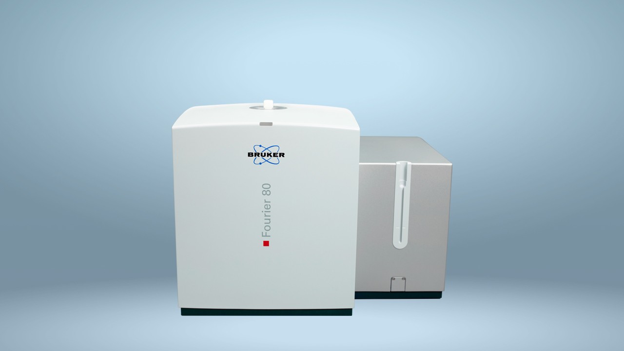
This application can also be used with our benchtop NMR system, the Fourier 80
![1H NMR spectrum for ethanol, showing spin coupling (green H signal split into sub-peaks). (Image by T.vanschaik (Own work) [GFDL or CC BY-SA 3.0], via Wikimedia Commons.)](/es/resources/library/application-notes-mr/nmr-101/_jcr_content/root/contentpar/twocolumns_575878242/contentpar-2/image.coreimg.gif/1698829959795/1h-nmr-ethanol-coupling-shown.gif)