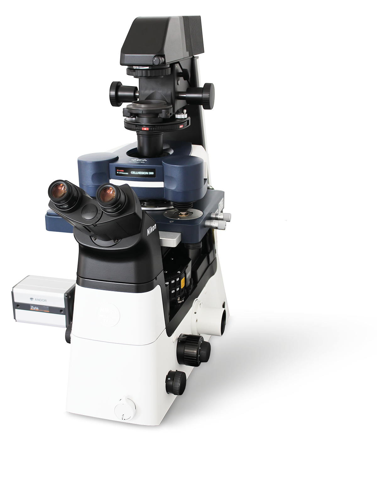
CellHesion® 300
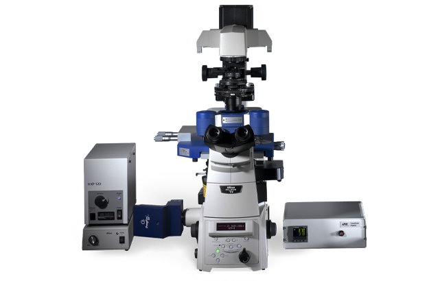
セルヘーション® 200
CellHesion® 200 は、細胞細胞と細胞基板の相互作用を測定するための統合システムです。このシステムは、外部の機械的ストレスに対する細胞の弾力性と細胞応答を定量化したり、組織をマッピングしたりするためにも使用できます。
細胞/組織力学と細胞接着の定量的な結果
力分光法データの品質は、分析を成功させるために不可欠です。最も低い電子騒音の床および最も堅い機械設計は必須である。装置の最高の正確さおよび安定性は例外的な性能の統合された容量性位置センサーによって保障される。最小ドリフトの場合、システム設計は対称です。
力曲線の最小変動の検出のためにJPKは、感度を改善し、ノイズレベルを損なうことなく検出帯域幅を増加させました。高いサンプリングレートと力曲線あたりのデータポイントの事実上無制限の数は、このユニークな楽器を完了します。
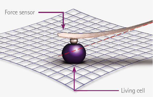
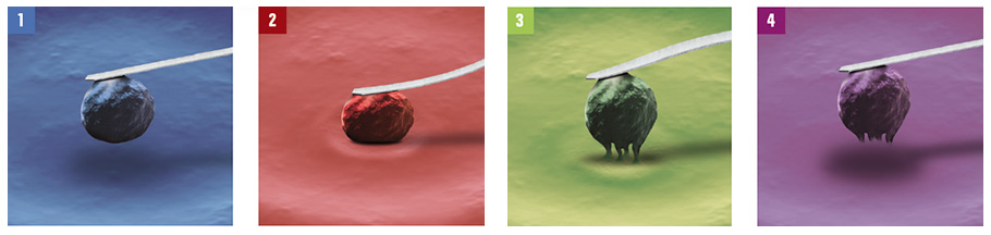
細胞接着操作原理
1. 単一の生細胞は、光学制御下でカンチレバーセンサー(例えば、フィブロネクチンコーティングを介して)に化学的に結合される。
この細胞は、結合ターゲット(分子層、インプラント表面、単一細胞、コンフルエント単層)との接触に定義された力を有する(スライド、カバースリップまたはペトリ皿)。
3. ユーザー定義反応時間の後、カンチレバー上の細胞は、ピエゾアクチュエータを介して垂直方向(z軸)にカンチレバーを引き込んで基質細胞から分離される。
4. セルは、ターゲットに付着した場合、表面から取り除こうとする試みに抵抗します。したがって、カンチレバーは顕著に曲がり、検出器によって測定される。
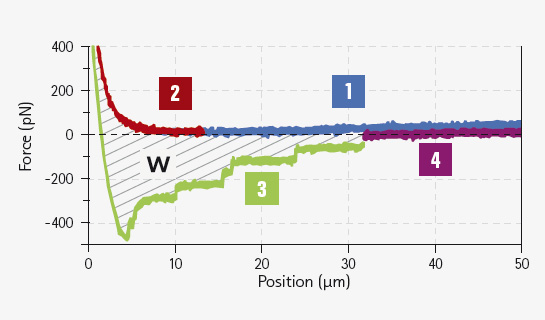
なぜなら、物理的には、片持ちは葉ばねであり、実際の接着力とエネルギーは測定された曲げから導き出すことができるからである。これにより、接着に寄与する単一分子結合事象の同定が可能となる。実験は、同じ細胞、異なるセル、異なるターゲット、および異なる条件で何度も繰り返され、統計的に関連する情報を得ることができます。
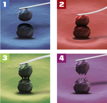
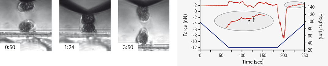
細胞基板接着原理
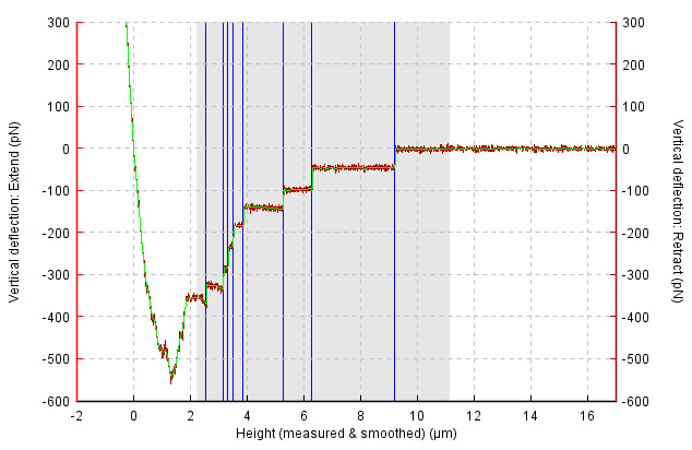
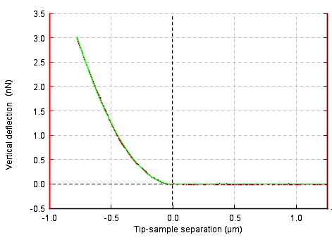
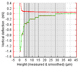
単一の測定サイクルの結果は、単一分子事象、「除去作業」W、テザー形成、最大接着力および粘弾性パラメータを決定することを可能にする力対距離曲線である。
光学統合のために作られる
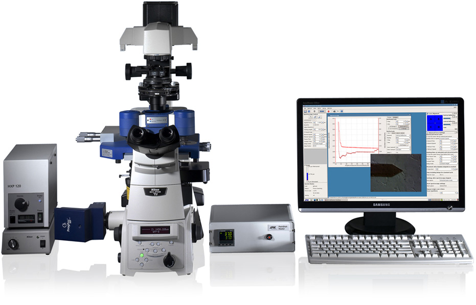
CellHesion® 200 をカール ツァイス、ライカ、ニコン、オリンパスなどの大手メーカーの反転した研究顕微鏡に簡単かつ完全に統合することで、強力な組み合わせになります。蛍光、共焦点顕微鏡、超分解能顕微鏡(STED、STORM/PALM)などの技術は、セルヘージオン®システムと同時に使用できます。
TIRF、CLSM、FRAP、Ca2+イメージングなどの蛍光技術を用いて細胞Hesionと並行して観察すると®接着過程や細胞骨格動態に関与する分子機構に関する知見が得られます。DICや位相コントラストなどの光透過照明技術のモードはすべて同時に使用できます。これは、細胞を力センサー(片持ちレバー)に移す場合や、検体の状態を確認する際の重要な特徴です。これらの光学的方法によって導出された構造情報は、CellHesion®200が電動式精密ステージまたは精密マッピングステージアドオンと共に使用される場合、DirectOverlay™ソフトウェア機能を介した力の測定によって決定される機能データと重ね合わせることができます。
細胞およびティッシュのための完全な環境
CellHesion® 200 は、生細胞を扱う特殊な要件に合わせて作られています。ガラス底または丸いカバーリップの有無にかかわらず35mmペトリ皿のような標準的な基質の使用は細胞の栽培および取扱いが容易になる。
15°Cから60°Cまでの温度制御、CO2制御用の流体交換およびポートは、JPK PetriDishヒーター™またはJPK BioCell™カバースリップ用に設計された統合されています。オプションとして、CellHesion® 200 は既存のインキュベーターに統合することができます (モデルを求める)。
サンプルと接触しているセットアップのすべての部分は殺菌することができる。CellHesion® 200 は、大きな細胞を処理し、さらによく接着する細胞を基質から分離するのに十分な垂直軸移動範囲を提供します。
アプリケーションの例
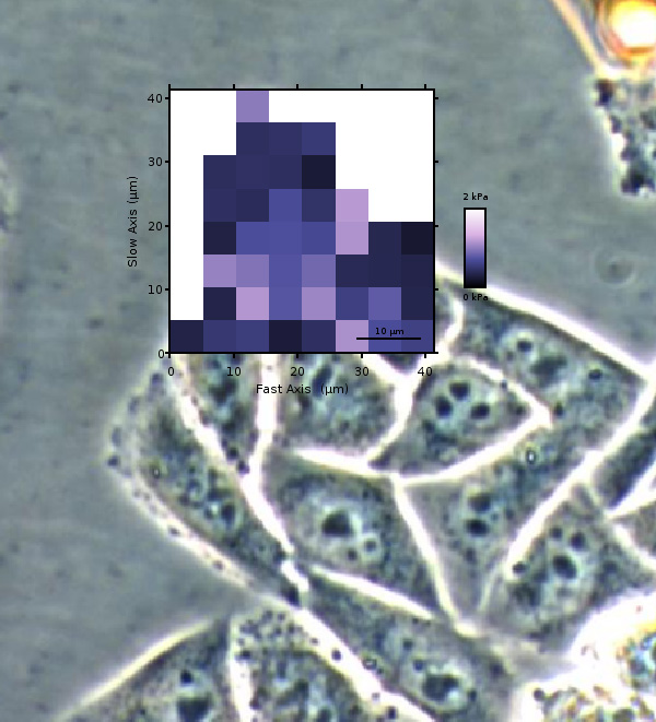
- 単一細胞から基質および組織への剛性と弾性マッピング
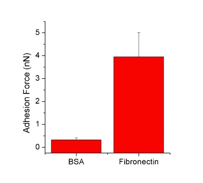
- 細胞細胞と細胞基質の相互作用
- 細胞接着とテザー形成
- 構造化基質、微小球、細胞に対する広範囲にわたるサンプル特性の自動マッピング。新しいハイブリッドステージなど™
- 生体材料研究、バイオファウリング、バイオセンサー、カプセル
- インプラントコーティングと細胞バイオチップ
- 微生物学・ウイルス研究への応用
- ドラッグデリバリー機構などの薬学的研究
- 繊維、コーティング、または空気または液体の粉末に関する食品、紙、繊維産業での用途
- 受容体/リガンド、抗体/抗原などの結合研究
- 機能化されたサーフェスのテスト
主な機能
- 細胞または組織の力学と接着実験のための革新的なプラットフォーム
- 細胞/細胞または細胞/基質相互作用、細胞弾性、テザー形成、接着性、細胞応答の特性評価
- 単一分子から細胞全体、組織、オルガネラ、胚までの定量的測定
- 構造化基質、微小球、細胞に対する広範囲にわたるサンプル特性の自動マッピング。新しいハイブリッドステージなど™
- 完璧なワークフローのための実験プランナー™直感的なユーザーインターフェイス
- DIC、位相コントラスト、蛍光、共焦点顕微鏡、超分解能、TIRF、フレット、FLIMなどの高度な光学イメージングと統合
- 幅広いモードとアクセサリ
セルヘーション®200データギャラリー
BrukerのBioAfMは生命科学および生物物理学の研究者が細胞の力学および付着、メカノ生物学、細胞細胞および細胞表面相互作用、細胞の動態および細胞形態の分野で彼らの調査をさらに促進することを可能にする。これらのアプリケーションのいくつかを示す画像のギャラリーを収集しました。
仕様
- 単一分子から細胞全体、組織までの測定に関する細胞接着/細胞力学研究のための革新的なプラットフォーム
- 複合実験用の逆顕微鏡との互換性、例えば、ツァイスアクシオオブザーバー、アクシコートA1、ニコンティ/TE 2000、ニコンティ2、オリンパスIXシリーズ、ライカDMiシリーズ
- DIC、位相コントラスト、蛍光、共焦点技術(TIRF、FRAP、LSMなど)などの主要な光顕微鏡技術に対応
- ツァイスアクシオズームV.16、ライカZ16アポア、ライカM205FA、オリンパスMVX10などの直立したマクロスコープとステレオ顕微鏡と互換性があります
- 強力な波形表面のためのz方向の自動範囲適応
- 高速容量性センサーフィードバックを通して閉ループ制御を備えた>110μmの移動範囲のカンチレバーセンサーリフティングシステム
- 電動精密ステージを装備し、特に組織探査用、20×20mm範囲内
- JPKバイオ™セルを使用したカバースリップ上のテンペチュア制御、灌流およびガスフロー(CO2)、またはウィルコ、BD、WPIのガラス底の有無にかかわらず35mmペトリ皿を持つネイティブ環境でのリビングセル研究™
- サンプルに接触したすべての部品は、きれいに簡単です
- 標準インキュベーターとの互換性(モデル指定)
- JPKソフトウェアバッチ処理によるセルラー相互作用パラメータのハイスループット判定
- 一分子事象、除去作業、最大接着力、結合解除イベント数、ヤング率などの粘弾性パラメータ(1つのシステム)
市場で最も幅広いアクセサリー
光学システム/付属品、電気化学ソリューション、電気サンプル特性評価、環境制御オプション、ソフトウェアモジュール、温度制御、音響および振動分離ソリューションなど。Brukerは、サンプル条件を制御し、実験を成功させるために適切なアクセサリを提供します。
最近のバイオAFMウェビナーを見る
当社の Webinar は、ベスト プラクティスをカバーし、新製品を導入し、難しい質問に対する迅速なソリューションを提供し、新しいアプリケーション、モード、またはテクニックのアイデアを提供します。