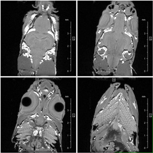
Anatomy and Morphology of Zebra Fish
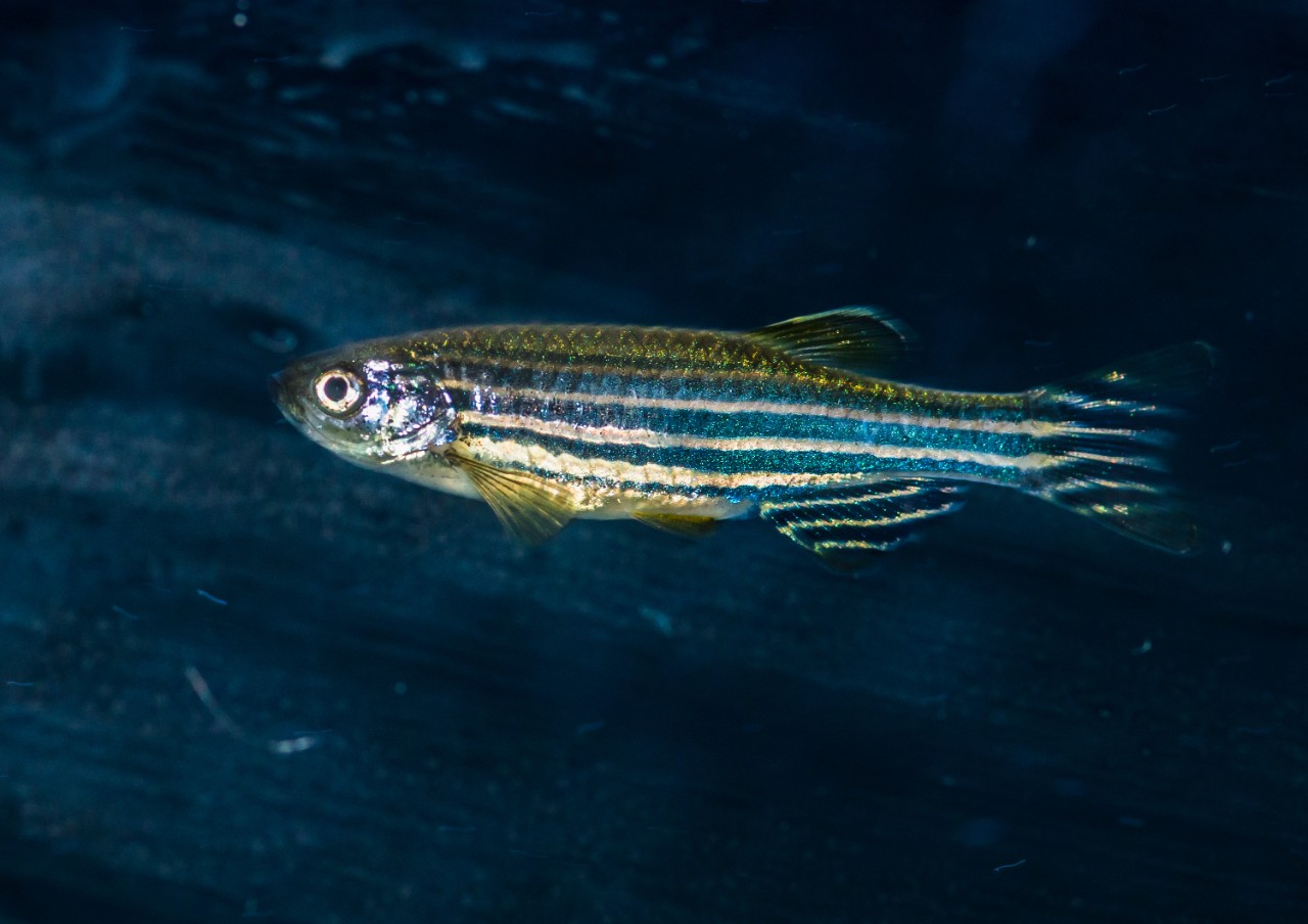
The zebra fish is one of the workhorses for the biogenetic research, due to its genetic structure similarity to humans. The fish is up to 4 cm long, and has a high reproduction and growth rate which makes it an ideal model organism for research on human diseases.
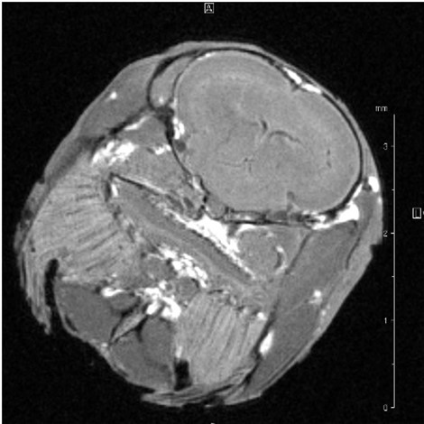
Non-invasive Magnetic Resonance Microscopy enables key insight into the zebra fish’s metabolism and morphology. Providing the researcher with a real-time tool for high sensitivity and resolution images and maps to monitor the organism’s development, for example when exposed to different environmental conditions, over and an extended period of time.
Magnetic Resonance Microscopy systems combine top of the line NMR systems with field strength up to 25.9 T with the state-of-the art microimaging accessory. Together with our wide range of room temperature probes which support sample diameters from 1 mm to 66 mm, and CryoProbes, the Magnetic Resonance Microscopy systems provide the ideal platform for your research.
References: Kabli, Samira, A. Alia, Herman P. Spaink, Fons J. Verbeek, and Huub J.M. De Groot. 2006. “Magnetic Resonance Microscopy of the Adult Zebrafish.” Zebrafish 3 (4): 431–39. https://doi.org/10.1089/zeb.2006.3.431.