Multi-modal PET drives interdisciplinary preclinical imaging
Multi-modal PET drives interdisciplinary preclinical imaging
Sonica Van Wyk, Market Product Manager, Nuclear Molecular Imaging, Bruker BioSpin
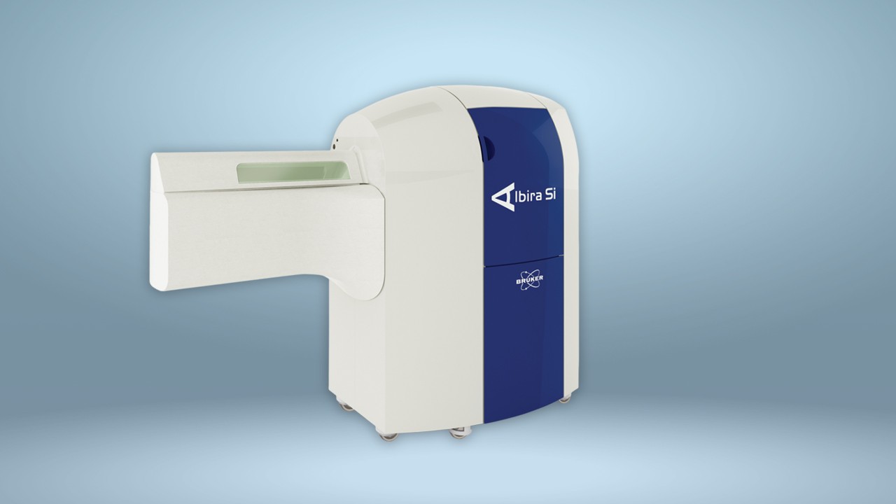
Tomography is an imaging technique used across a wide variety of fields, ranging from radiology and nuclear medicine, to geophysics and materials science. It provides threedimensional information about a subject based on its sections or projections, and common examples include X-ray computed tomography (CT), positron emission tomography (PET) and single-photon emission computed tomography (SPECT). CT scanning provides information on the anatomy of the subject, while PET provides functional imaging which shows the spatial distribution of biomolecular activity in the body. PET was developed as a technology for both clinical diagnostic
and preclinical purposes in the 1950s, the scope of which was expanded by the development of radiopharmaceuticals – a group of pharmaceutical drugs which emit radiation and commonly includes radiotracers.
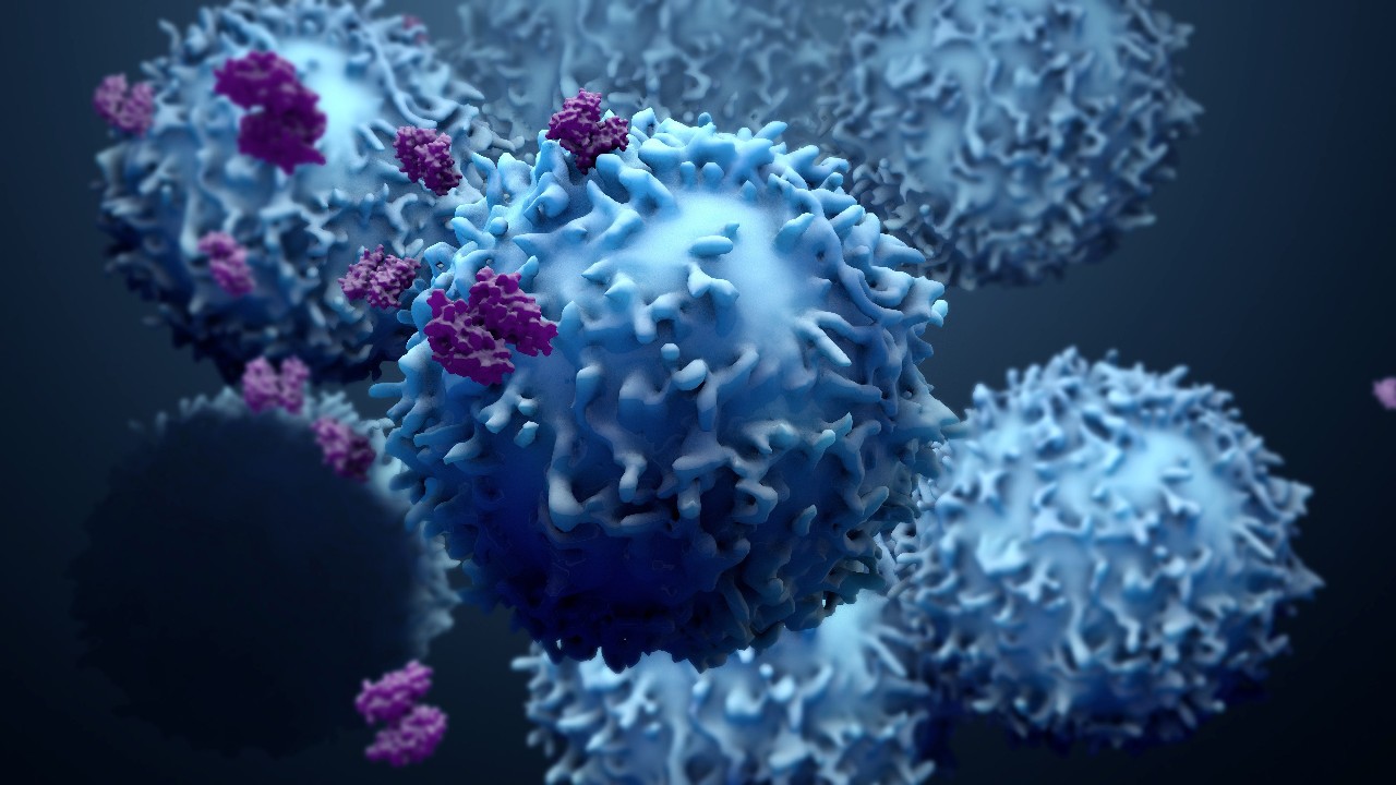
Preclinical imaging (PCI) plays a vital role in understanding the biological processes behind disease states at the organ, tissue, cell and molecular level. Elucidating how the body responds to physiological or environmental change is important in the search for therapeutic agents to fight disease. PCI is also critical to the evaluation of new treatment effectiveness and safety, by informing researchers of drug distribution patterns in tissues. PET in preclinical studies enables users to conduct repeat experiments on the same animal subjects, providing strong statistically valuable data and therefore reducing the number of animals required for a study. For this reason, it has becoming increasingly important to use non-invasive in vivo imaging techniques to optimize the use of each animal used.
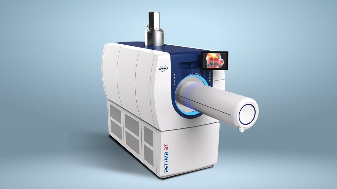
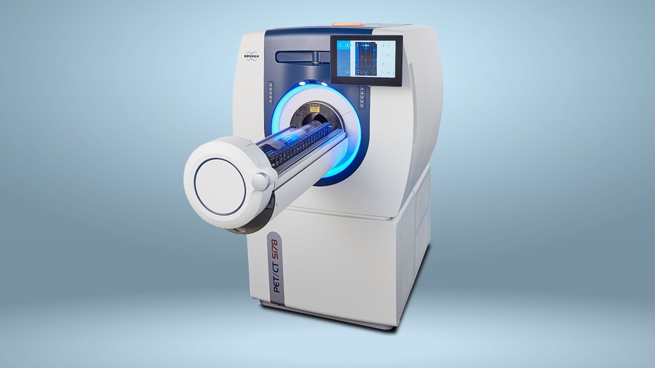
Multi-modal tomographs, such as PET/CT, allow the correlation of the functional imaging obtained using PET with the anatomic imaging obtained with CT scanning. PET can also be combined with other technologies, such as magnetic resonance imaging (MRI), to bring functional imaging together with soft tissue morphological imaging. PET/MR is gaining ground in preclinical imaging applications, as it offers superior soft tissue contrast, imaging without the CT’s ionizing radiation risk, and multiparametric data.
Download the full application note: