Previously Unknown Conformation of TNFα Identified Using DEER
“The new structure offers opportunities to modulate the biological activity of tumor necrosis factor α”
Tumor necrosis factor α (TNFα) is a pluripotent cytokine that mediates a diverse range of biologic effects. Although the precise function of TNFα is unclear, blood levels are increased in patients with a chronic inflammatory disease, such as rheumatoid arthritis and Crohn’s disease, suggesting that it plays a role in the pathogenesis of such disorders.
An inflammatory function of TNFα is supported by clinical data showing the benefit of anti-TNFα agents in the treatment of a range of inflammatory diseases. The biologic anti-TNFα drugs, such as infliximab, adalimumab, etanercept, and certolizumab pegol, have demonstrated significant efficacy in chronic inflammatory diseases, including Crohn’s disease, rheumatoid arthritis and psoriasis.
Although biologic anti-TNF treatments have revolutionised the treatment of inflammatory diseases, they are associated with injection-site reactions and increased risk of developing serious infections, including tuberculosis, and lymphoma1.
Consequently, there has been much research into the possibility of achieving similar efficacy using small molecules, which may increase the therapeutic safety window with regard to increased risk of infection and cancer.
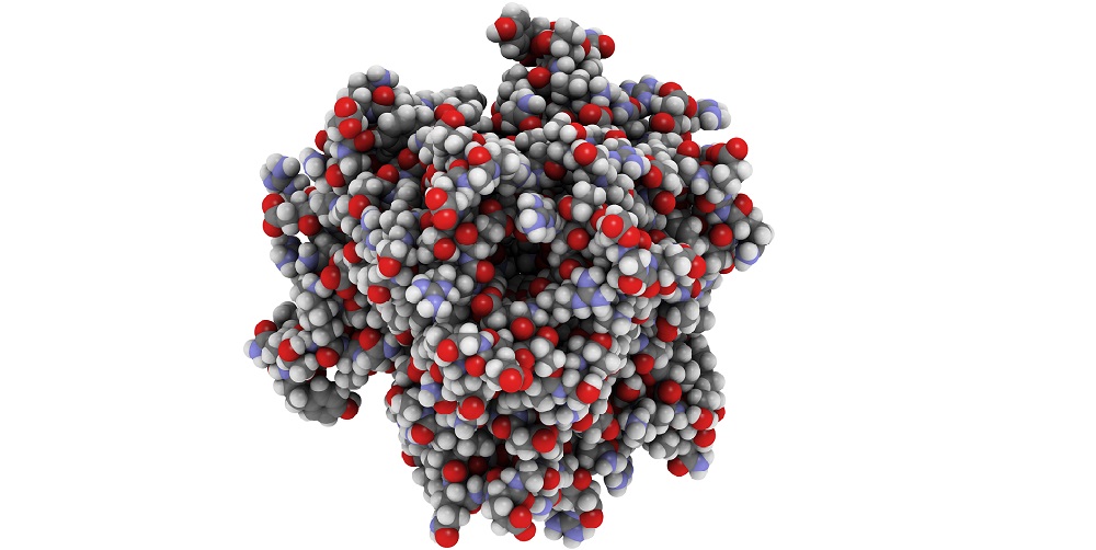
A key part of this research is structural analysis of TNFα to try and identify possibilities for modifying its activity using small molecules. TNFα comprises three monomeric units that associate to form a trimer that binds TNFα receptors.
Immunoenzymatic assays have demonstrated that TNFα can readily switch between the inactive monomer and the active trimer2. This conversion is concentration-dependent with the trimeric form appearing transiently in regions of high concentration.
It appears that inhibitors of TNFα work by causing a conformational change that results in dissociation of the active trimer to the inactive monomers3. Researchers were therefore keen to identify whether there was a particular inactive configuration of the TNFα trimer that could be stabilized to reduce the level of TNFα activity.
Determination of the structural conformation of the trimer immediately before dissociation to its constituent monomers would allow the search for suitable small molecules that may stabilize the distorted, inactive trimer to begin.
Double electron-electron resonance (DEER), a technique well suited to the elucidation of protomer movement within a trimer, has recently been used in conjunction with site-directed spin labeling to study natural conformations of the human TNFα trimer4.
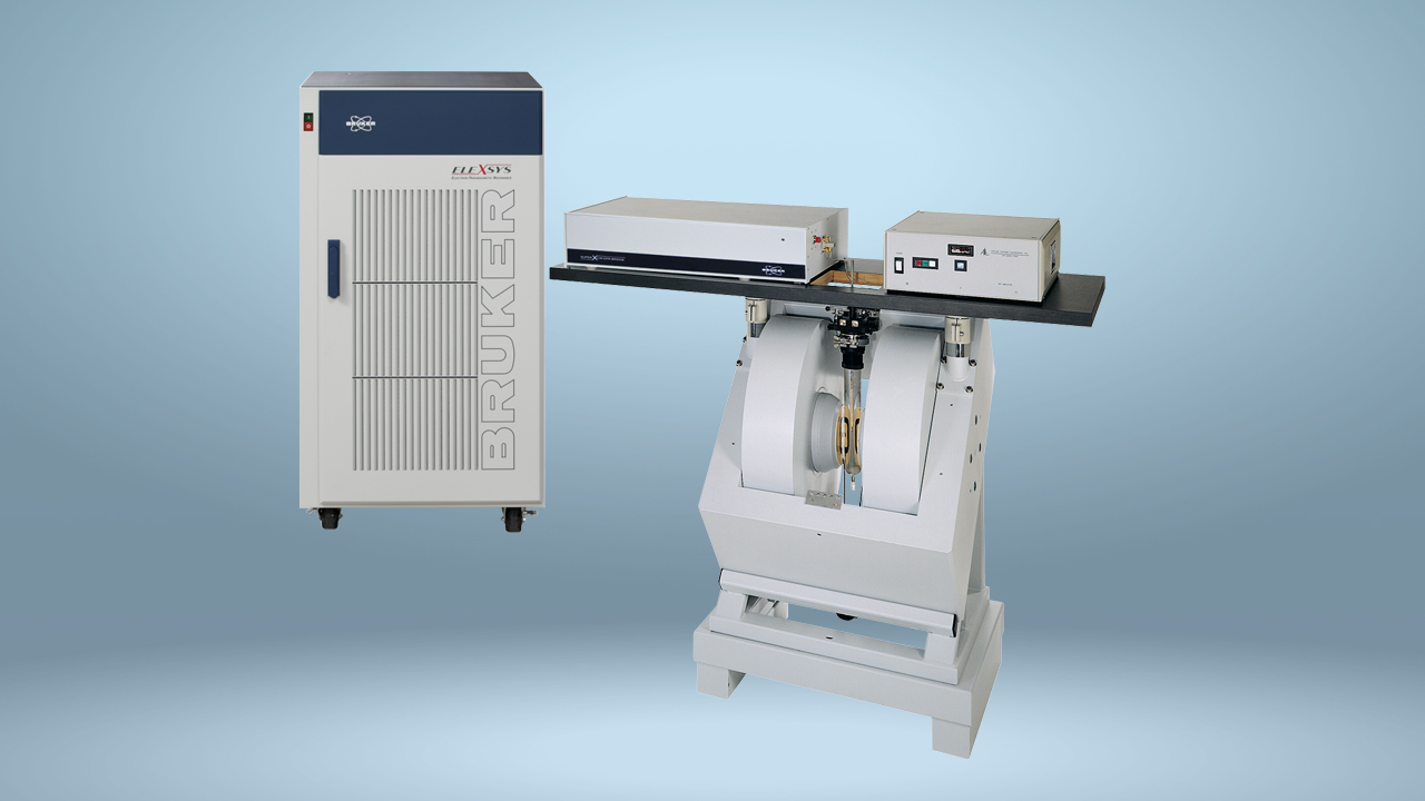
A Bruker BioSpin E580 spectrometer was used for the DEER analysis. All pulses in Q-band ELDOR were performed with a Bruker SpinJet arbitrary waveform generator and a Bruker dielectric TE01δ resonator. Continuous-wave electron paramagnetic resonance was conducted using a Bruker BioSpin SHQE-W TE011-mode cylindrical resonator.
A previously unreported, predeoligomerization conformation of the trimer was identified. In this newly identified structural conformation of the TNFα trimer, one of the monomers is rotated and tilted outward. It is this movement of the monomer that leads to the dissociation of the trimer, preventing binding to the TNFα receptors.
The new structure offers opportunities to modulate the biological activity of TNFα by stabilizing the distorted trimer, which cannot bind the TNF receptor and so is not effective, with small molecules.
References
- Ding T and Deighton C. Complications of Anti-TNF Therapies. Future Rheumatol. 2007;2(6):587‑597.
- Corti A, et al. 1992. Oligomeric tumour necrosis factor a slowly converts into inactive forms at bioactive levels. Biochem. J. 1992;284:905–910.
- Alzani R, et al. Mechanism of suramin-induced deoligomerization of tumor necrosis factor a. Biochemistry. 1995. 34:6344–6350.
- Carrington B, et al. Natural Conformational Sampling of Human TNFα Visualized by Double Electron-Electron Resonance. Biophysical Journal 2017;113:371–380.