Highlighting MRI as a Technique for Preclinical Neuroscience Research
How can neuroscience research help us to find new insights into brain function?
We can use magnetic resonance imaging (MRI) to provide 2- or 3-D images of the brain for the study of its anatomy, function, or molecular processes... or a combination of all three. The nice thing about MRI is that a researcher can chose whether the focus should be anatomical with a little bit of functional, for example, or, whether it should be molecular.
What information can in vivo neuroimaging give us about brain function and metabolism?
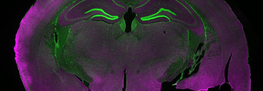
Using a technique called diffusion MRI, we can track axons directions throughout the brain and create connectivity maps of the brain, in a non-invasive and non-destructive manner.
On the functional side, we have a number of options. Functional MRI (fMRI) enables us to watch the brain while it thinks. This technique is a clinical standard and for more than a decade, we have been able to apply it to animals including rats and mice. Contrast agents are not required; we just monitor subtle signal changes due to the conversion of oxy- and deoxy- haemoglobin that enable us to clearly detect brain activity.
In addition, we are able to monitor changes in the cerebral blood flow, which is an important marker. In stroke research, we can see affected brain regions, probably with a higher precision than with most other non-destructive methods.
To investigate metabolites in vivo spectroscopy can be used. Using this, we can obtain chemical "fingerprints" of brain regions. The size of these regions are typically some millimeters cubed and the fingerprint that enables us to identify and quantify some dozens of metalbolites in that volume. These include major neurotransmitters and molecules involved in the energy pathway of the brain.
Why isn't MRI always a familiar technique amongst biologists?
MRI is typically not included in the biology curriculum. MDs receive a basic training in the background of MRI, and even much more if they eventually become radiologists. Biologists, however, are first introduced to these scanners when they use it as a technique to address a biological question. I studied both biology and chemistry, and in chemistry, I learned all the basics of NMR and MRI. If I had studied just biology, however, I would have never learned about the great possibilities of MRI.
Every biologist learns how to handle an optical microscope but, unless their university has a pre-clinical MRI scanner, they'll not be familiar with MRI technology. Bruker's MRI application experts have put their knowledge into pre-optimized protocols. Even users with minimal MRI background can quickly answer their biological questions.
Please outline the use and importance of MRI & PET/MRI in fundamental neuroscience research.
PET lacks anatomical information. Generally speaking, with PET, you track wherever your tracer goes in the body, and what you end up seeing are the only areas where the functionalized tracer is located.
If you use PET alone, you cannot really be sure where these active areas are within the body because you have no anatomical reference. With the PET/MRI combination, you can overly. The colored PET image on the gray scale high-resolution MRI image and then you can see with high precision exactly where your tracer is located.
The importance and the beauty of the combination of PET and MRI is that you can perform both at the same time and get great soft tissue contrast out of your MRI.
What are the benefits of these imaging techniques over other methods?
Aside from being non-destructive, there is the fact that we can get more information using less animals.
You can achieve greater statistical relevance because you can use the scanner to study the same animal repeatedly over a period of weeks or months. Animals are not sacrificed after each study time point, instead, we scan the whole cohort and get all the information from all of the animals. Each animal serves as its own control. This reduces the biological scatter which is an inherent problem of a lot of preclinical studies. I think this is a huge benefit that is often overlooked.
Are findings from preclinical research fully translatable to clinical setting? Can preclinical imaging of the brain do things which are not possible clinically?
They are translatable, yes. The animals that are imaged with PET and MRI experience the same procudeures that patients do in a hospital with clinical instruments. Of course, there are benefits of preclinical imaging, such as testing novel disease treatments before they go to the clinic. You can also use knockout models to look at the mechanisms of disease progression.
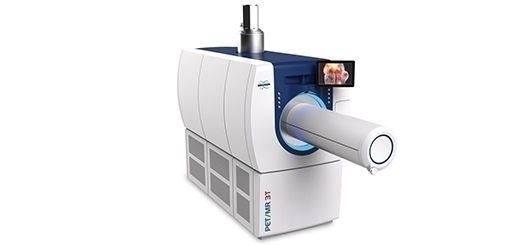
Please give an introduction to the Bruker instrumentation that is used for preclinical neuroscience research.
We have a line of pre-clinical MRI scanners, which we introduced more than 40 years ago. We are the leaders in the market. Bruker's preclinical MRI scanners are called BioSpecs and there are various different versions. You can choose from a range of magnetic fields. The higher the magnetic field, the better the images in general. The other thing you have to choose is what we call the bore - the little tunnel inside the magnet in which the animal lays during the examination. Small-bore scanners can only hold a mouse, while other larger ones can holdrats or even larger animals.
Our PET scanners also have small tunnels for rats and mice. We also offer a combination of PET and MRI. In one PET/MR design, the PET tunnel is in front of the MRI tunnel, so the two machines are adjacent and the animal is on a kind of a monorail, which goes to the PET tunnel first for a quick scan. Then you move it forward by about 20 inches to position it in the MRI scanner, where you can perform your MRI scanning.
There is an alternative PET/MR setup in which the small PET ring fits directly into the MRI tunnel and enables the animal to go right into the center of the MRI scanner, which is also the center of the PET scanner. You can then scan simultaneously.
Which preclinical disease models has this instrumentation been used to investigate? Has it been able to identify any potential treatments?
Well, it's basically endless and ranges from models of Alzheimer's disease and Parkinson's disease to models of memory, aging, and cognitive decline, to name but a few. The instrumentation is also used in stroke research. If we artificially induce stroke in a rodent, we can quantify the affected brain area probably better than with any other method that does not involve dissection of the brain. Many pharma companies use Bruker scanners in drug discovery and development.

Support

Service and Life Cycle Support
Bruker’s commitment to provide customers with unparalleled help throughout the buying cycle, from initial inquiry to evaluation, installation, and the lifetime of the instrument is now characterized by the LabScape service concept.
LabScape Maintenance Agreements, On-Site On-Demand and Enhance Your Lab are designed to offer a new approach to maintenance and service for the modern laboratory