ROS Detection Alzheimer's
The Effect of UV Exposure on AMyloid Plaque Aggregation in Alzheimer’s Disease
Alzheimer’s disease (AD) involves the progressive deterioration of mental faculties including cognition, rational thinking, and memory. At times, this disease can become debilitating and can increase the risk for other health issues. Poor memory, for example, can cause an AD patient to lose a significant amount of weight because this person may have forgotten to eat at regular mealtimes. Those affected by AD progress in their disease differently, and patients with AD also vary in regard to the level of disease severity. Medications are available to help slow the progression of AD; however, a cure for the disease remains elusive.
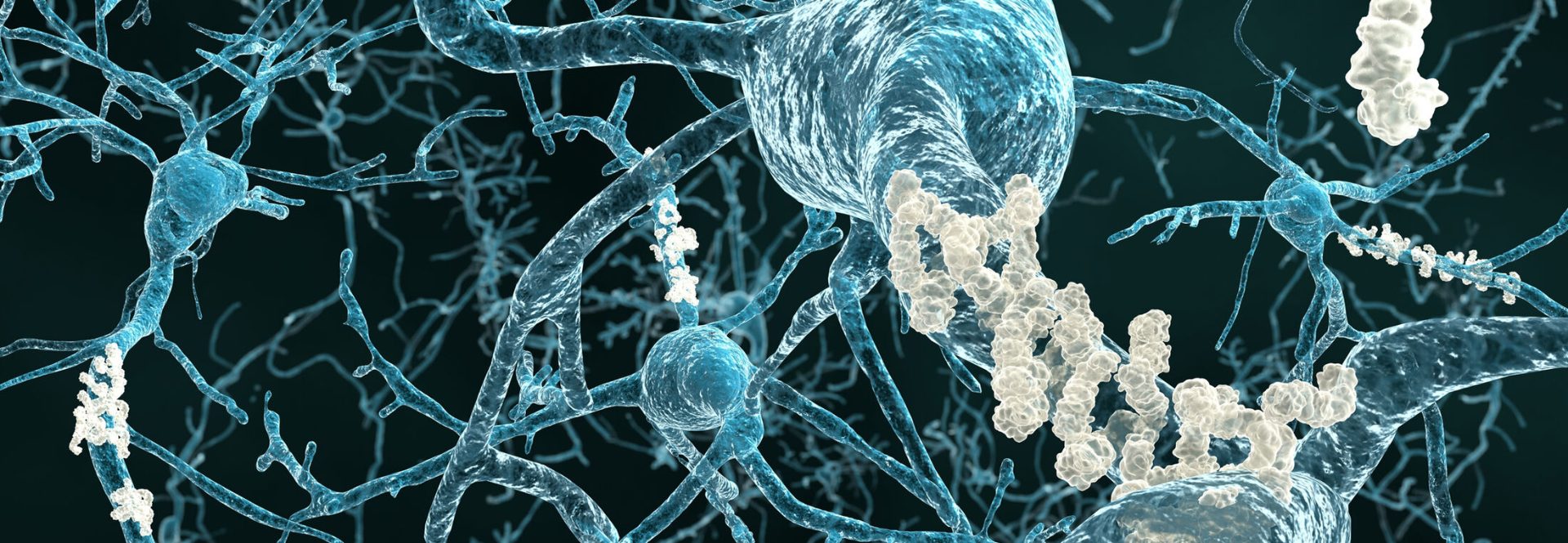
Currently, many researchers are focusing their efforts on amyloid plaques which are typically found as deposits in the brains of AD patients. The main component of these plaques is Aβ peptide, and aggregation of these peptides are what cause the plaque formation.
AD Pathogenesis and Reactive Oxygen Species (ROS)
Research has consistently found a correlation between AD pathogenesis and the generation of reactive oxygen species (ROS) and soluble Aβ oligomers. Additional research has also found that within many AD patients, high concentrations of zinc (Zn) and copper (Cu) exist within the amyloid deposits. The interaction between Cu2+-Aβ can result in significant brain toxicity for AD patients, a discovery that has recently facilitated more attention toward chelation therapy of Cu2+ from Cu2+-Aβ aggregates as an approach to AD. Specifically, researchers believe the Cu2+-Aβ interactions are reversible and Cu2+ binding to Aβ peptides have the potential to be regulated by chelation.
Catalytic photooxygenation has previously been discovered to reduce the effect of neurotoxicity and aggregation of amyloid β (Aβ) peptides. In a study by Xiongwei Dong et al, the effect of ultraviolet (UV) exposure ≤400 nm on chelator-treated and -untreated Cu-bound aggregates was examined.
Researchers found that UV light markedly enhanced the cytotoxicity of Cu2+ free and -bound aggregates. Upon recognizing this finding, researchers further examined whether this effect truly was due to the UV exposure or the chelation treatment.
Using Electron Paramagnetic Resonance (EPR) to Evaluate Free Radical Content
Two groups were analyzed using a Bruker X-band EPR spectrometer system. EPR is used to evaluate a sample’s free radical content.
The two groups were as follows:
- In the presence of FC-11-1 or FC-11, Cu2+ bound and -free Aβ 42 aggregates at 10 μ M that were either exposed to 1500 Lux UV light for 4 hours or incubated in darkness.
- A control group also exposed to 1500 Lux UV light for 4 hours or incubated in darkness, consisting of 10 μ M FC-11 or FC-11-1, 10 μ M cupric sulfate, and 10 μ M FC-11-Cu2+ or FC-11-1-Cu2+ in a phosphate buffer solution.
In this experiment, EPR was specifically utilized to identify the generation of ROS by aggregates exposed to UV light and aggregates incubated in darkness in the presence of FC-11-1 or FC-11.
Further examination showed that controlled UV light exposure caused Cu2+-free and -bound Aβ42 aggregate dissociation and produced ROS. In addition, researchers reported that the chelators used (FC-11 and FC-1) enhanced cytotoxicity of the Cu2+-bound Aβ40 aggregates due to the treated aggregates dissociation into soluble oligomers. While there is no conclusive decision on what these results mean for the treatment of AD, there is hope that this study can shed new light on the effects of UV exposure for patients with progressing AD.