Activation of Hippocampal CA3 Reduces fMRI-BOLD Response in the Brain
Functional magnetic resonance imaging (fMRI) is widely used for mapping neuronal networks and identifying synchronized activities within the brain. It is also used to study functionally connected regions in the brain and to test the assumption that as one area fuels activity in a different area via direct connections, the intensity of fMRI signals in the regions will develop similarly.
fMRI studies often involve the electrical stimulation of i.e. the forepaw, detecting where this stimulation is registered in the brain via modifications in blood oxygen level dependent (BOLD) signal.
Combined stimulations of the medial prefrontal cortex as well as the anterior cingulum regions may result in BOLD responses in common neuronal network regions. An additive response may occur when a target region receives excitatory (glutamatergic) inputs from two different brain regions.
In a study published in PLoS One by Scherf T et al, fMRI was used to detect networks within the brain that are “activated” via electrical stimulation.1 This electrical stimulation either occurs through the ventral tegmental area (VTA) or hippocampal CA3.
An animal model, including Wistar rats, was used for this study. All electrophysiological responses were evoked and recorded using a stimulus generator, and signals were transferred to an analogue-to-digital interface.
Researchers in this study stimulated either the hippocampus (the hippocampal CA3, specifically) or the VTA (neurons close to the midline on the midbrain “floor”). They found that this stimulation resulted in significant responses of BOLD in common brain structures, including the left and right hippocampus and the septum. Other BOLD responses were also observed in the striatum and medial prefrontal cortex during VTA stimulation.
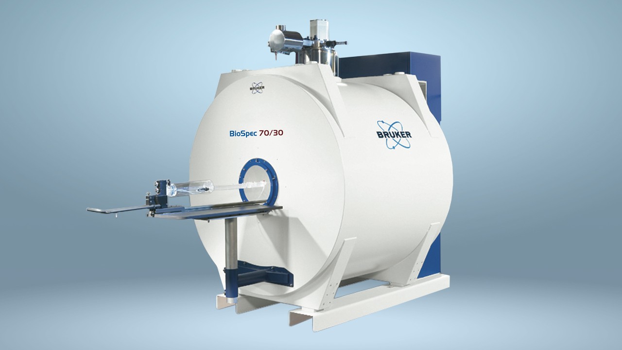
Researchers relied on the Bruker BioSpec 47/20 for the study.
The fMRI and electrophysiological experiments were combined into three stimulation blocks, with each block containing similar, if not identical, stimulation trains. The total duration for the combined session was around 35 minutes.
Further findings revealed that electrical stimulation of the VTA resulted in significant dopamine release in target regions of the dopaminergic mesolimbic pathway, which connects the VTA with the nucleus accumbens. This release, in turn, caused a BOLD response. The researchers also showed that with the use of low-frequency 2 Hz pulses for stimulating the left hippocampus, a reduction in BOLD responses in the anterior cingulum regions and medial prefrontal cortex occurred.
An additional incoming excitatory (glutamatergic) input was also observed, which was a somewhat surprising finding. This input can degrade a BOLD response formation in the anterior cingulum and medial prefrontal cortex regions, exerting possible consequences for the understanding of functional connectivities resulting from corresponding modifications of BOLD signal intensities.
In combination with these results, the study also observed that the VTA and left hippocampus were either separately or concurrently stimulated. Each brain structure extends to varying and common target regions, and it’s the combined stimulation that may cause different activation patterns when compared with only one structure receiving this stimulation.
References
Scherf T., Angenstein F. Hippocampal CA3 activation alleviates fMRI-BOLD responses in the rat prefrontal cortex induced by electrical VTA stimulation. PLoS One. 2017;12(2):e0172926.