EPR 101
Free Radicals Revealed
Electron paramagnetic resonance (EPR) spectroscopy, also known as electron spin resonance (ESR) offers insights into otherwise invisible phenomena by detecting atoms and molecules containing unpaired electrons (i.e. they are paramagnetic). It distinguishes itself from other spectroscopic techniques in that only EPR unambiguously detects these species, thus eliminating false positives.
EPR is a highly sensitive and specific technique, that enables static and dynamic studies of materials, chemical samples, and biological systems.
People use EPR spectroscopy to:
- Detect, identify, and quantify free radicals
- Study molecular structures, geometry, and dynamics
- Observe labeled species in situ in biological systems
- Understand redox processes, reaction kinetics, and more
EPR can detect any species with paramagnetic electrons, including organic and inorganic radicals, transition metal complexes, metalloproteins, biradicals, and more.
Free radicals occur quite frequently in nature due to one electron transfer reactions. Transition metal ions are often paramagnetic.
EPR Strenghts
EPR spectroscopy provides direct measurements of the number and type of nuclei coupled to unpaired electrons. An excellent complement to NMR, it brings unique capabilities to any analytical lab:
- Sensitivity – up to 1000 times more sensitive than NMR
- Specificity – only molecular regions containing unpaired electrons detected
- Fast time resolution – monitor even short-lived species over time
- Non-destructive measurement – leaves samples intact for further analysis
- Quantitative analysis
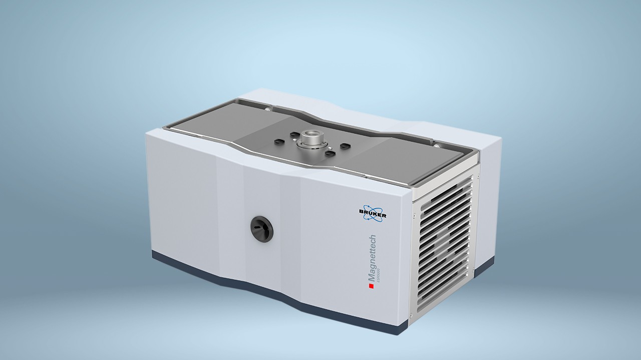
First commercialized in the 1950s, EPR has become much more accessible, thanks in part to the development of modern radar technology. Benchtop EPR systems now offer greatly enhanced ease-of-use, reduced the cost of ownership, and advanced capabilities in a minimal footprint.
Inside EPR
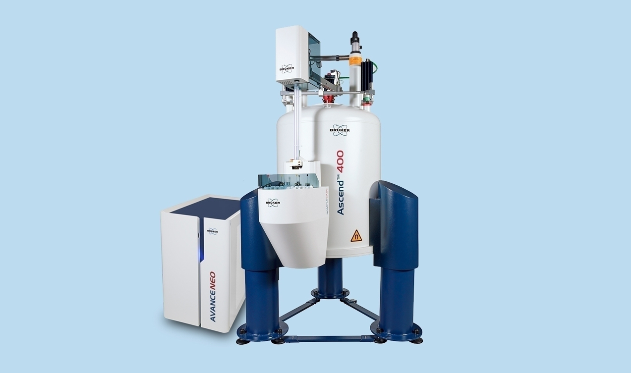
EPR spectroscopy is analogous to NMR spectroscopy, with a central difference. EPR generates measurements by probing the magnetic properties of unpaired electrons, rather than atomic nuclei as in NMR. (See NMR101.)
As a charged particle in motion, every electron has a magnetic moment. Placed within a magnetic field, the unpaired electrons in a sample align their magnetic moments with and against the magnetic field. The external magnet’s field is linearly swept while exposing the sample to a fixed frequency of microwave irradiation. The condition where the magnetic field and the microwave frequency are “just right” to produce an EPR resonance (or absorption) is known as the resonance condition in which the electron moment changes its orientation with respect to the magnetic field.
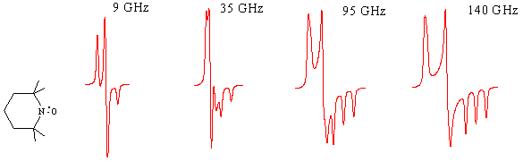
Specific paramagnetic species absorb energy at characteristic “resonance” frequencies, which vary with the strength of the magnetic field. Electromagnets make it relatively easy to sweep across a range of strengths, so most EPR experiments hold frequency constant while measuring energy absorbed as a function of magnetic field strength. The resulting absorbance spectrum reveals details about the presence and environments of free electrons in the sample.
Here’s a brief overview of the process:
Sample introduction:
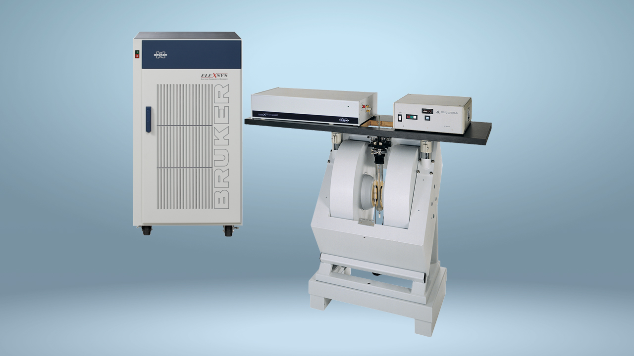
After minimal preparation, the sample is placed inside a probehead often called a cavity or resonator, which in turn sits in an electromagnet. EPR is often performed at low temperatures, using liquid helium or nitrogen as coolant, to capture fleeting responses.
Data acquisition:
The resonator is a physical structure that resonates (stores and also concentrates the microwave energy) at the wavelength of the microwaves, much like an organ pipe resonates with sound waves. When the sample absorbs the microwaves while appropriate microwave frequency and magnetic field are applied, microwaves are reflected back from the cavity and our EPR signal is detected.
Data interpretation:
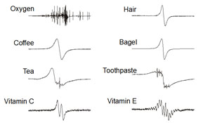
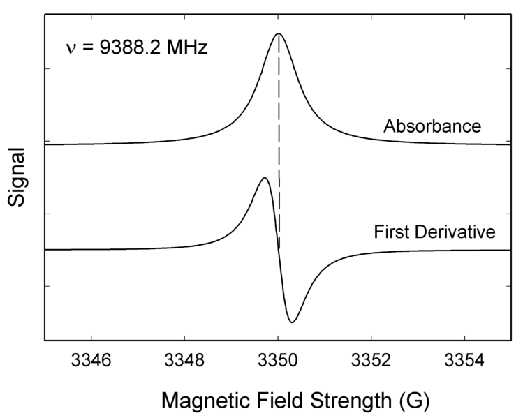
Unlike many spectroscopic techniques the absorption of the microwaves is presented as a first derivative spectrum. This is because EPR uses field modulation and lock-in detection techniques to achieve ultimate detection sensitivity as well as offering superior resolution of signals.
If free electrons in a sample had only two possible states – such as “excited” (after absorbing a photon) and “relaxed” – then the EPR spectrum would show a single zero crossing, like the idealized example above. In reality, multiple electromagnetic influences can create a variety of possible energy states, with corresponding lines on the EPR spectrum. Their pattern reveals details about the species present. Among the most important EPR parameters, the “g factor” reflects interactions between an electron’s orbital and spin angular momentum. Electron interactions with magnetic nuclei, called hyperfine interactions, provide valuable information about molecular structure and identity.
EPR Applications
EPR’s unique views into paramagnetic species make it valuable to research, development, and quality control efforts across fields and industries. For example:
Biology and Medicine
- Studying metalloproteins in metabolic function
- Monitoring formation and reactivity of reactive oxygen species (free radicals)
- Using spin-labels to study membrane proteins and dynamics of protein-lipid interactions
Materials and Physical Sciences
- Identifying crystal defects in metal oxides
- Developing and testing semiconductors
Chemical and Petrochemical Industries
- Studying reaction kinetics, catalysis, photochemistry, and more
- Real-time monitoring of crude oil for free-radical-containing Asphaltenes
Food and Beverage Quality
- Tracking product shelf life
- Assessing oxidative-, temperature- and photo- stability
- Identifying free radicals in irradiated foods
Learn More
See the following articles for more detailed examples of EPR in action.
- Food Irradiation Control Using EPR
- Freshly Brewed Research Reveals Coffee’s Antioxidant Power
- Rapid, automated analysis for optimizing your beer’s shelf life
Continue exploring the possibilities of EPR with these free webinars.
- A Wider World of EPR – At Your Benchtop
- Bring EPR into the Classroom