Sedimentology
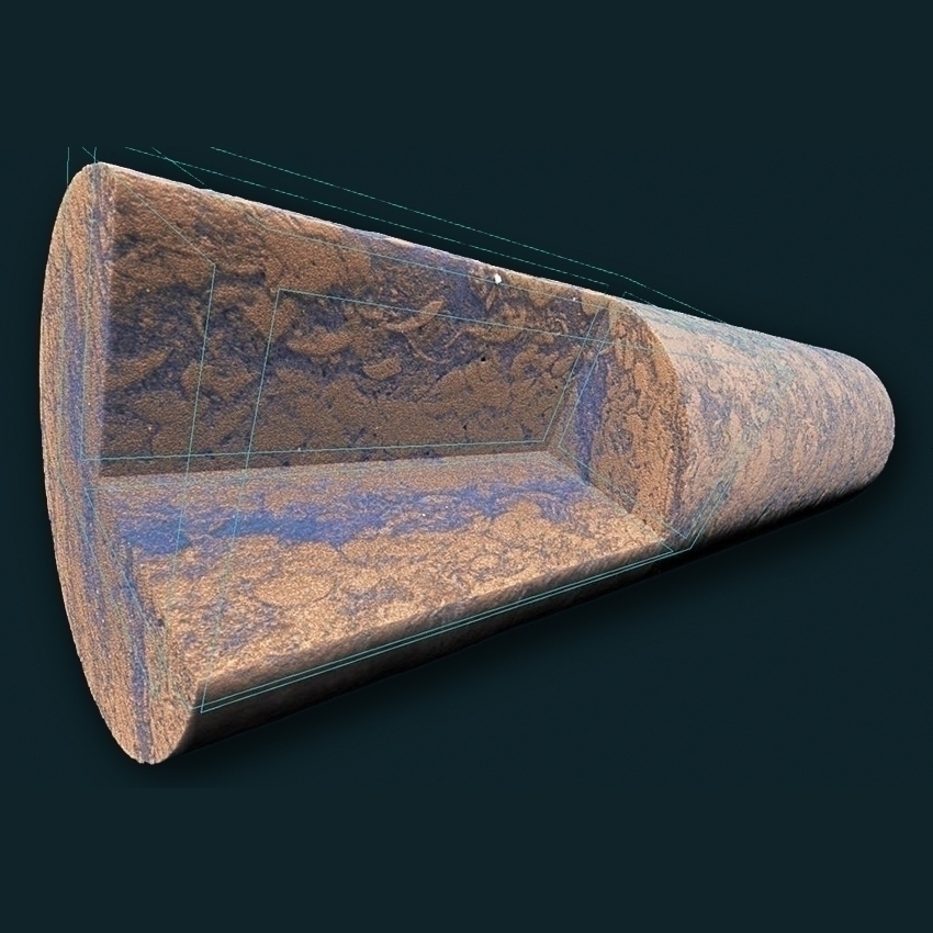
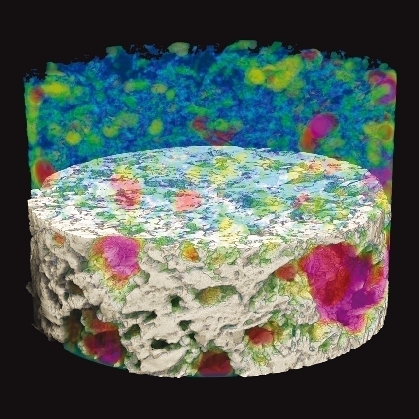
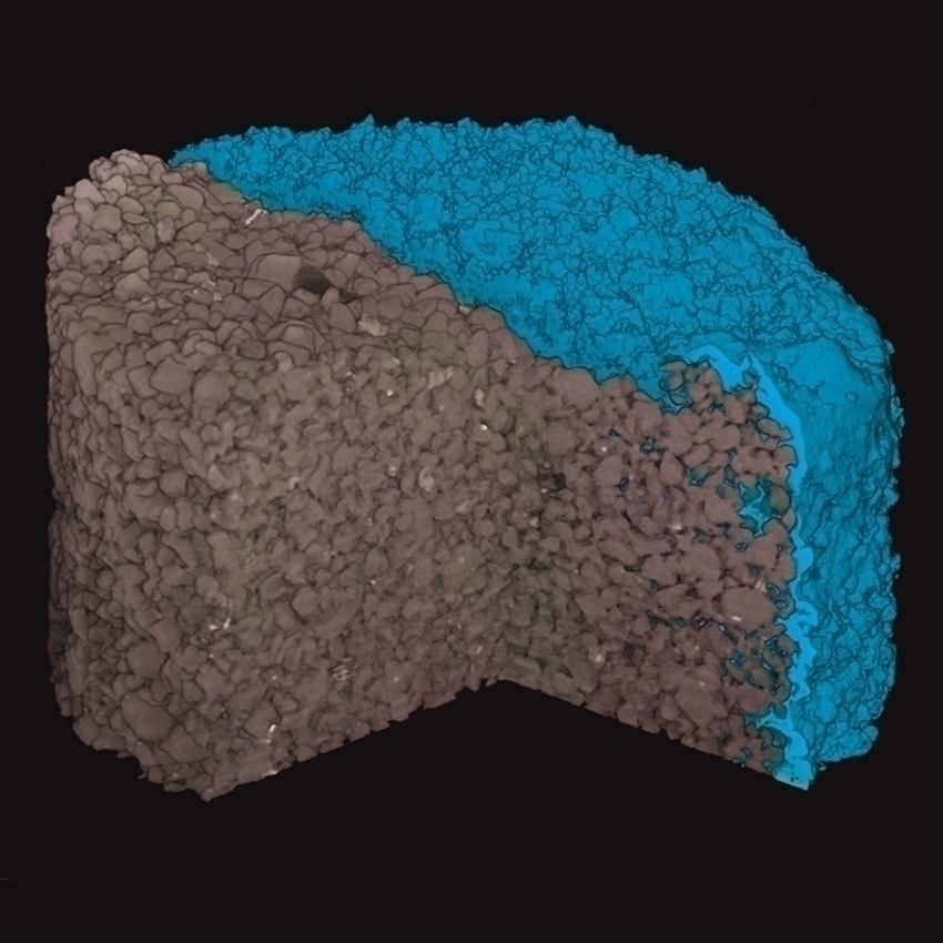
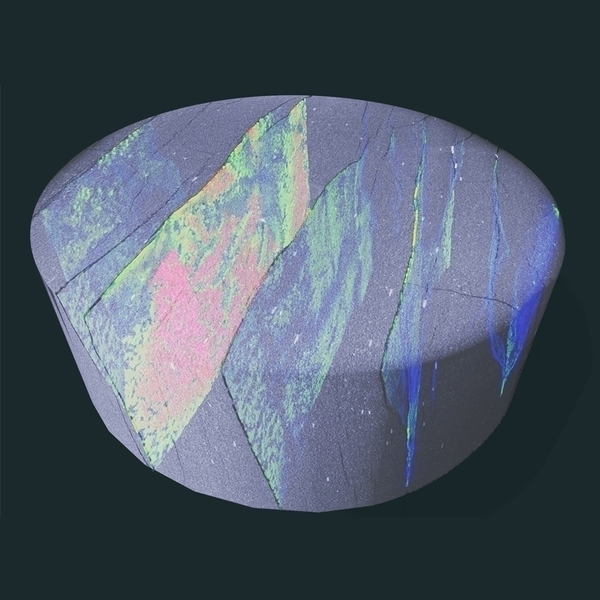
3D X-Ray Microscopy (XRM) uses micro-computed tomography (micro-CT) for high-resolution 3D characterization of sedimentary rocks. XRM is a non-destructive 3D imaging analysis technique for samples from 1 mm to 20 cm. The system rotates the sample in a chamber and collects thousands of radiographs at different angles. The radiographs are backprojected into a virtual space with voxels constructing a volume with X-ray signal attenuation represented in 3D space. The attenuation is related to density and atomic number and can be used to understand porosity, mineral distribution, and structure. Voxel resolution can be as low as 0.4 µm depending on sample size; for example, a 1 µm resolution can be expected for a 2 mm sample.
Common applications for XRM in sedimentology include 3D characterization of:
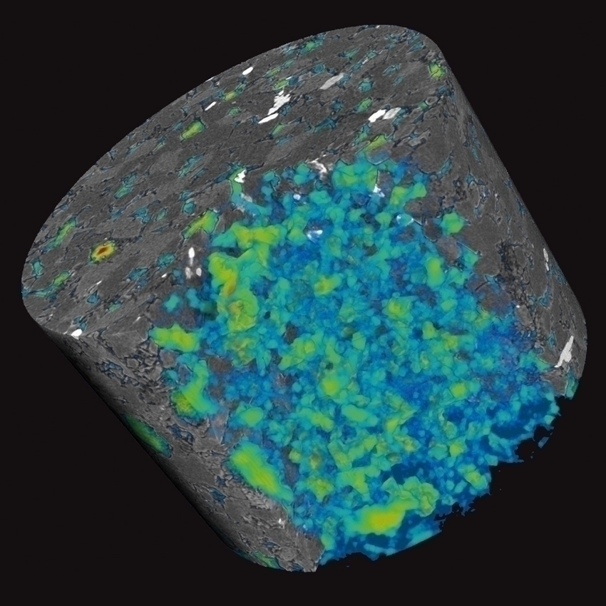
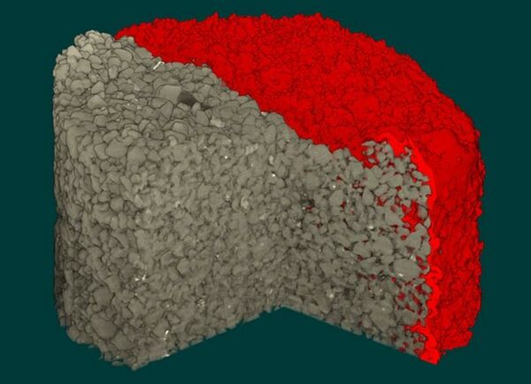
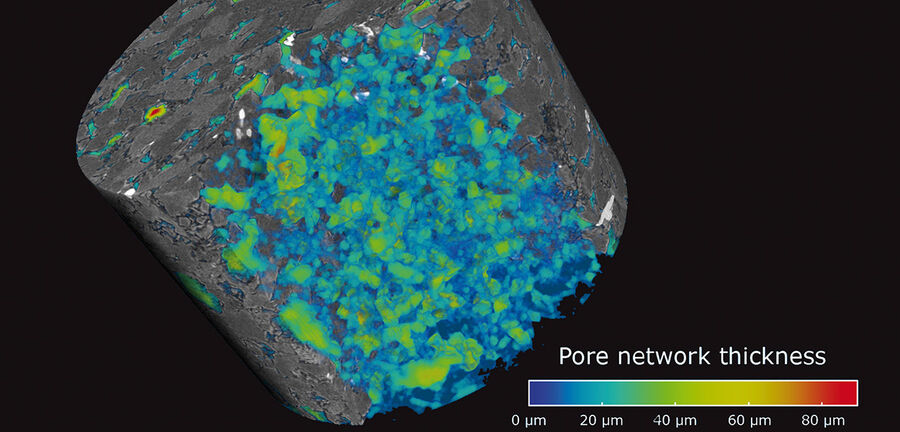
- Porosity and pore network properties measured for reservoir characterization and fluid flow modeling including permeability modeling, multiphase flow properties calculations, capillary pressure curves and resistivity index.
- Mineral phase segmentation, which allows volumetric calculations, particularly when combined with optical microcopy, micro-XRF with the M4 TORNADO AMICS, or automated mineralogy with AMICS for SEM.
- Sediment texture including grain sphericity, shape and orientation enabling grain size analysis.
- Soft sediment deformation, structural deformation and sedimentary structures, enabling process reconstruction and calculation of original flow conditions without the bias of outcrop or thin section.
Key References
Ketcham & Carlson
Computers & Geosciences, 27, 381–400, 2001
Applications of X-ray computed tomography in the Geosciences
Mees, Swennen, Van Geet, Jacobs
Geological society, London, Special Publications Volume 215, 2003
Wildenschild & Sheppard
Advances in Water Resources, 51, 217–246, 2013
Cnudde & Boone
Earth-Science Reviews, 123, 1–17, 2013
Noiriel
Reviews in Mineralogy and Geochemistry, 80, 247-285, 2015