The Application of NanoWizard ULTRA Speed AFM to Study Dynamics of Living KPG7 Fibroblasts
KEYWORDS: Dynamic Cellular Processes; Fast-Scanning AFM; Fluorescence Microscopy; High-Speed AFM; Fibroblasts; Live Cell Research
Dynamic measurement of live cells at near-physiological liquid conditions requires faster image acquisition than is typically achievable with conventional AFM imaging. Moreover, working with these often soft, topographically inhomogeneous materials is made more challenging by their weak and rapidly changing sample signals.
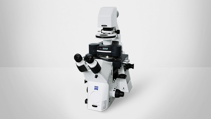
Nevertheless, imaging soft biological matter and rapidly changing, very active living cells is a fundamental part of studying dynamic cellular interactions. These events are typically impossible to observe with conventional AFM, but can be captured using the fast-scanning NanoWizard ULTRA Speed AFM due to its high spatiotemporal resolution, improved feedback response, and other leading-edge enhancements.
In this application note, we demonstrate these capabilities. The NanoWizard ULTRA Speed AFM is successfully used to monitor cytoskeletal reorganization and cell membrane protrusion events in living KPG7 fibroblasts in nearly physiological buffer conditions.
Readers can expect to learn about:
- AFM-based investigation of challenging biological/soft materials and systems;
- How fast-scanning AFM can be used and combined with advanced optical techniques to gain new information about cellular interactions that were previously impossible to observe; and
- The collection of dynamic measurements on living cells and direct observation of cell membrane dynamics with the NanoWizard ULTRA Speed AFM.