Bruker Atomic Force Microscopy Solutions Enable Rapid Imaging of Native Infectious and Inactivated SARS-CoV-2 Viral Particles
The global research effort surrounding the novel coronavirus (SARS-CoV-2) responsible for Coronavirus Disease 2019 (COVID-19) has progressed at an unprecedented rate. Given that the virus is classified as a hazard group 3 pathogen, however, only laboratories certified as level 3 biosafety (BSL3) facilities are able to study SARS-CoV-2 in its active state. To facilitate ongoing endeavors to better understand the virus’ structure and function, there remains a need for effective inactivation methods that allow labs with lower biosafety clearance to study SARS-CoV-2 and thus contribute to the growing knowledge base.
Viral particles can be inactivated by a variety of methods, including the application of heat, alcohol, peroxide, radiation, fixatives, or detergents, but, to date, few have demonstrated the ability to maintain SARS-CoV-2 native morphology.1,2 A recent study by researchers at the University of Montpellier, France, investigated the ability of formaldehyde (FA) to inactivate SARS-CoV-2 viral particles while preserving their morphology.3
AFM is a unique tool that enables imaging of biological samples in their native liquid environment at a molecular scale. Using this method, samples do not need to be fixed, coated, or stained, allowing scientists to image both native and inactivated viral particles in buffer, at high resolution and in three dimensions (3D). In this study, the AFM measurements were performed directly in the BSL3 laboratory using the NanoWizard IV bio-atomic force microscope (bio-AFM) from JPK Instruments-Bruker.
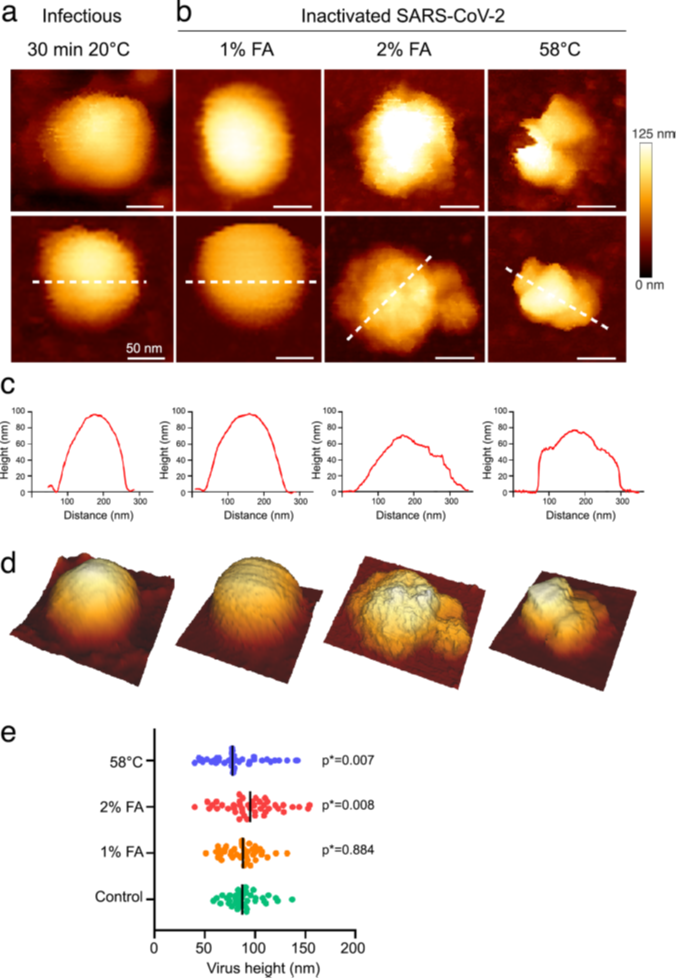
The group examined and compared virus inactivation using FA and heat to evaluate the extent to which each method could keep SARS-CoV-2 structurally intact. Following inactivation treatment by either FA at four different concentrations (0.5, 1.0, 2.0, and 3.6%) or heat, viral particles underwent ultra-filtration and direct analysis by AFM to analyze nanoscale morphology. SARS-CoV-2 was successfully inactivated after incubation at 58°C for 30 mins, but AFM analysis showed severely damaged particles that had lost their spherical shape (Figure 1b).
All four FA concentrations showed complete SARS-CoV-2 inactivation (when incubated at 20°C for 30 mins), but viral particles were damaged at 2.0 and 3.6% FA (Figure 1c-e). The study also confirmed existing reports that the FA inactivation effect was temperature-dependent and significantly compromised at 4°C.
The inactivation methods described in this study can expand the number of potential laboratories that can carry out SARS-CoV-2 research outside a BSL3 setting, and high-resolution AFM has been shown to be a reliable tool for analyzing SARS-CoV-2 in buffer to provide fast, direct qualitative information on virus morphology. By maintaining viral morphology, scientists can study the structure and function of non-infectious particles and ultimately contribute to the ongoing efforts to better understand the virus’ mechanisms of infection and transmission, and potentially lead to the discovery of therapeutics.
Read the full paper: https://www.nature.com/articles/s41598-021-91371-4
For more information on AFM technology, please visit https://www.bruker.com/en/products-and-solutions/microscopes/bioafm.html
References:
- Patterson, E. I. et al. Methods of Inactivation of SARS-CoV-2 for downstream biological assays. J. Infect. Dis. 222, 1462–1467 (2020).
- Storm, N. et al. Rapid and complete inactivation of SARS-CoV-2 by ultraviolet-C irradiation. Sci. Rep. 10, 22421 (2020).
- Lyonnais, S. et al. Atomic force microscopy analysis of native infectious and inactivated SARS-CoV-2 virions. Scientific Reports. 11:11885 (2021).