IT
My Bruker
Contatta un esperto
Prodotti e soluzioni
Applicazioni
Servizi
News & Eventi
Chi Siamo
Lavora con noi
Utilizza almeno 2 caratteri (attualmente stai utilizzando 1 carattere).
Languages
Deutsch
English
Español
Français
Italiano
Polski
Português
Русский
中文
日本語
한국어
Multiphoton Microscopes
Multiphoton Microscope Webinars
Hear from researchers and experts on best practices, new techniques and discoveries, and ideas for new applications using two-photon microscopy.
Utilizza almeno 2 caratteri (attualmente stai utilizzando 1 carattere).
Multiphoton Microscopes
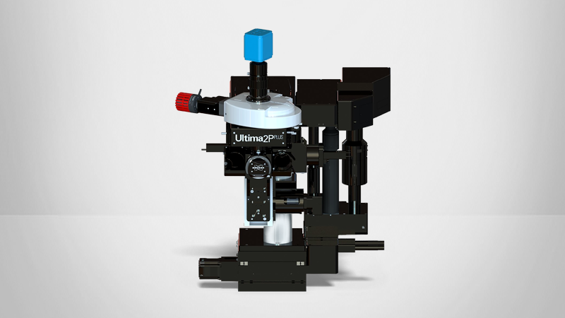
Ultima 2Pplus
A complete all-optical multiphoton workstation for imaging and optical manipulations
Leggi di più
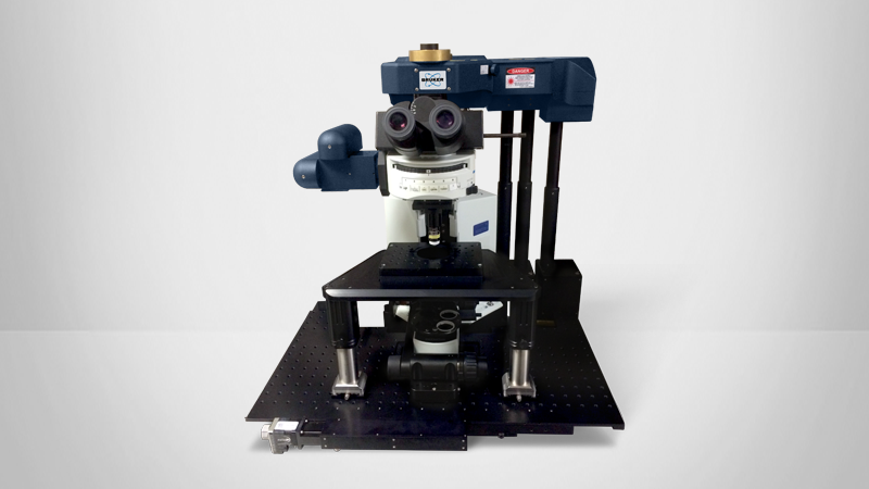
Ultima In Vitro
A timeless workhorse for simultaneous multiphoton imaging and photoactivation (a.k.a Prairie Scope)
Leggi di più
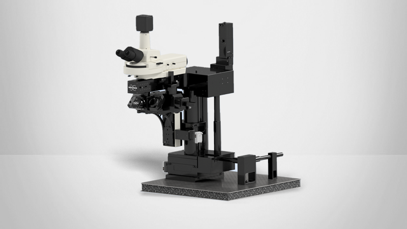
Ultima Investigator Plus
An accessible, large field-of-view multiphoton microscope that grows with your research
Leggi di più