Simple Minds, Sophisticated Analyses Illustrate Anesthesia-Specific Influence on Brain Connections
Why do we do the things we do, and what happens in our brains when we do those things? Untangling the complex functional connections in the human brain is a profound undertaking. How can you track intricate connections in an advanced being when there are so many variables? Add to that the gauze of anesthesia, and making sense of all those connections becomes nearly impossible. In some ways, turning to a simpler brain model might just make it possible to track brain functions and tweeze out connections without the noise of advanced minds. That simpler brain is present in the ubiquitous and ever-serving mouse.
Maybe seeing how brain networks are connected in those small brains under resting state functional magnetic resonance imaging (rs-fMRI) could illustrate just how brain regions connect and work. On top of that, given how frequently scientific studies focus on rodents under anesthesia, knowing which and how connections are altered by anesthesia would be useful in distinguishing the anesthesia-related effects from the changes in functional connectivity caused by disease, injury or treatments.
To show a more complete “brain signature” for specific anesthesia, researchers recently applied analyses techniques primarily used in human research to rs-fMRI data collected on mice. They used dual regression followed by graph theory-based network analysis to gain new knowledge about functional networks and brain systems of mice under anesthesia. The approach provided information on “brain-wide” networks as opposed to information just about isolated regions.
The results appear in Bukhari, Q., Schroeter, A., Cole, D. M., & Rudin, M. (2017). Resting State fMRI in Mice Reveals Anesthesia Specific Signatures of Brain Functional Networks and Their Interactions. Frontiers in Neural Circuits, 11, 5.
The team relied on data from an earlier study, Grandjean J., Schroeter A., Batata I., Rudin M. (2014). Optimization of anesthesia protocol for resting-state fMRI in mice based on differential effects of anesthetics on functional connectivity patterns. Neuroimage 102, 838–847. 10.1016/j.neuroimage.2014.08.043
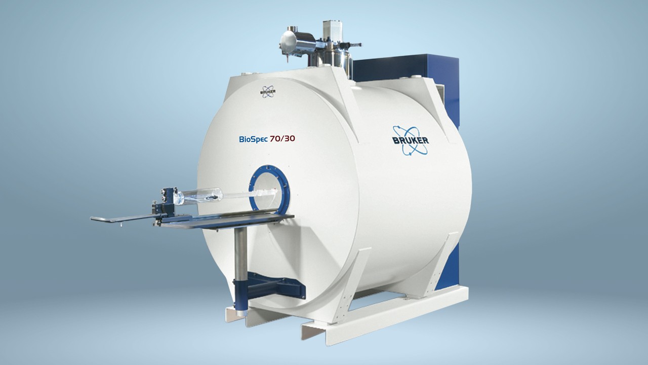
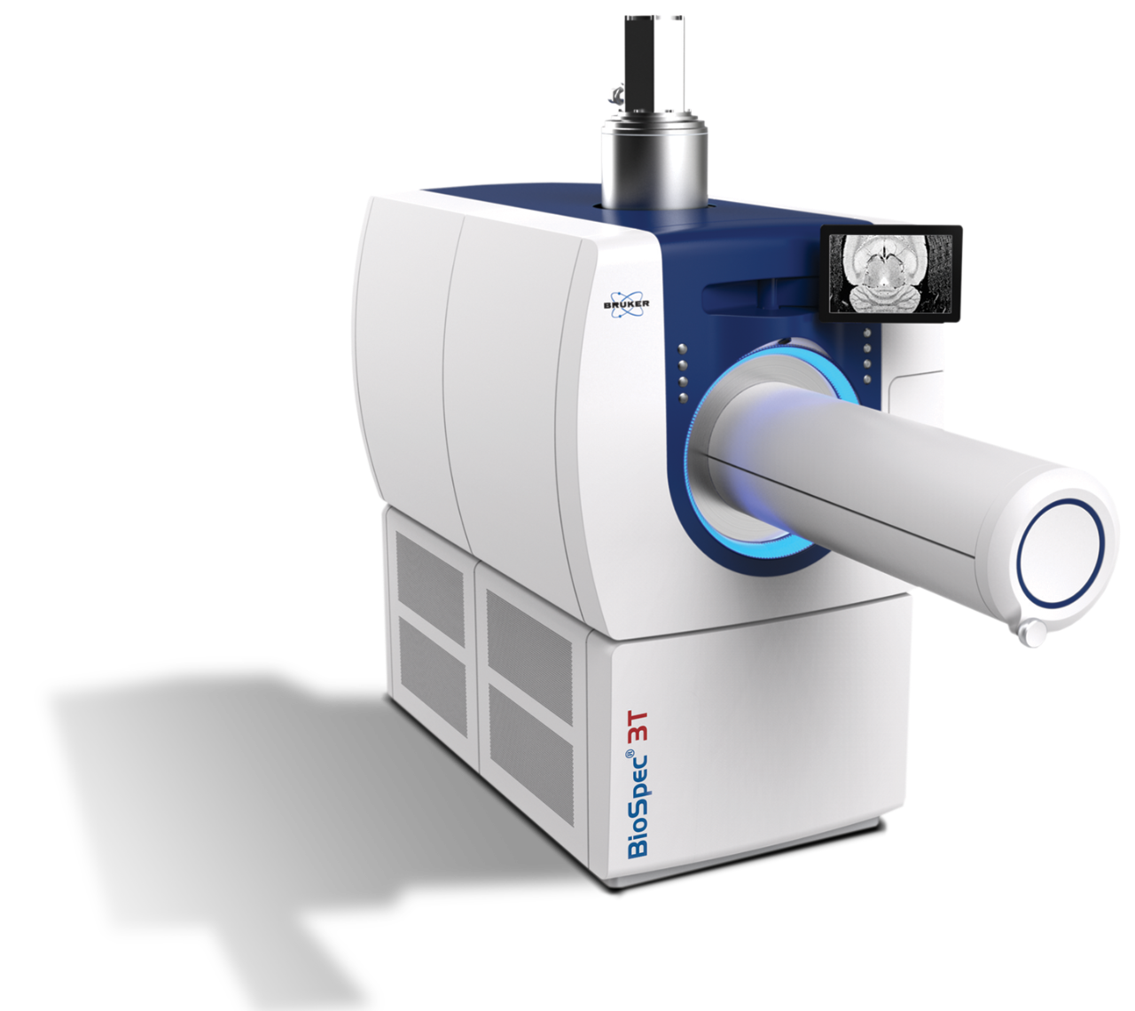
The rs-fMRI data were gathered using a Bruker Biospec 94/30 small animal MR system operating at 400 MHz (9.4 T) equipped with a four-element receive-only cryogenic phased array.
Making the most of rs-fMRI results through advanced data analyses
By using a combination of data analyses techniques, including independent component analysis (ICA), dual regression and network modeling, researchers identified five networks in the mouse brain where within- and between-network activity is altered in anesthetic-specific ways. Lateral cortical, associative cortical, default mode, subcortical and thalamic. Specifically, researchers looked at two common anesthetics, Isoflurane and medetomidine.
Mice receiving isoflurane primarily demonstrated interactions in intra- and inter cortical areas. However, mice on medetomidine mostly displayed subcortical functional connectivity, including between cortical and thalamic locales.
The researchers then considered whether combining isoflurane and medetomidine at lower doses could provide the necessary benefits with less of the clouding effects of each anesthesic separately given at full dose. That’s exactly what they found, and the complementary nature of the medicines made it easier to see functions and connections throughout the brain using rs-fMRI. The mice were effectively anesthetized and, “retained strong correlations both within cortical and subcortical structures, without the potential seizure-inducing effects of medetomidine, rendering this regimen an attractive anesthesia for rs-fMRI in mice.” In addition, this brain-wide approach showed how areas connect in a way that studies of isolated regions of the brain just can’t.
Detailed and comprehensive information on rodent brain function under anesthesia can be useful in selecting the most appropriate anesthesia, refining data interpretation and, potentially, translating that knowledge to human brain function.
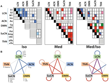
Figure: Illustrating functional networks and their interaction as derived from DR analysis
(A) Matrices displaying within- and between-network interactions under the various anesthesia regimens. Matrices have been structured according to the five functional networks identified. Colors indicate significant interactions observed under isoflurane (blue) or medetomidine anesthesia (red). For the group receiving the combination anesthesia, elements are indicated in different colors depending on whether they apparently arise from the isoflurane group (blue), from the medetomidine group (red), from both groups (blue/red) or were observed in the combination group exclusively (green). Positive correlations are indicated by dark colors, while light colors represent negative correlations. Gray blocks indicate the absence of any significant interaction. Significant group differences (with Bonferroni correction) in connectivity have been observed for all the interactions displayed.
(B) Within- and between-network interactions detected in isoflurane (blue lines), medetomidine (red lines) and medetomidine/ isoflurane anesthetized mice. In isoflurane/medetomidine anesthetized mice network interactions, blue lines represent interactions observed in mice under isoflurane anesthesia only, red under medetomidine only, and violet represents interactions observed for both anesthesia regimes. The width of the lines in the figure indicates the strength of the connectivity.
References
Bukhari, Q., Schroeter, A., Cole, D. M., & Rudin, M. (2017). Resting State fMRI in Mice Reveals Anesthesia Specific Signatures of Brain Functional Networks and Their Interactions. Frontiers in Neural Circuits, 11, 5. http://doi.org/10.3389/fncir.2017.00005
Grandjean J., Schroeter A., Batata I., Rudin M. (2014). Optimization of anesthesia protocol for resting-state fMRI in mice based on differential effects of anesthetics on functional connectivity patterns. Neuroimage 102, 838–847. 10.1016/j.neuroimage.2014.08.043
A comparative encyclopedia of DNA elements in the mouse genome. The ENCODE Project Consortium. A user’s guide to the encyclopedia of DNA elements (ENCODE). PLoS Biol. 9. e1001046 (2011)
Chang, C., Leopold, D. A., Schölvinck, M. L., Mandelkow, H., Picchioni, D., Liu, X., Ye, F.Q., Turchi, J., Duyn, J. H. (2016). Tracking brain arousal fluctuations with fMRI. Proceedings of the National Academy of Sciences of the United States of America, 113(16), 4518–4523. http://doi.org/10.1073/pnas.1520613113
Jonckers E., Shah D., Hamaide J., Verhoye M., Van der Linden A. (2015). The power of using functional fMRI on small rodents to study brain pharmacology and disease. Front. Pharmacol. 6:231. 10.3389/fphar.2015.00231
Pan W. J., Billings J. C., Grooms J. K., Shakil S., Keilholz S. D. (2015). Considerations for resting state functional MRI and functional connectivity studies in rodents. Front. Neurosci. 9:269. 10.3389/fnins.2015.00269