Torsional Resonance Dynamic Friction Microscopy (TR-DFM)
Nanoscale Characterization For Next-Generation 2D Materials
Expanding upon Bruker’s innovative Torsional Resonance Mode (TR-Mode™), Torsional Resonance Dynamic Friction Microscopy (TR-DFM) provides increased sensitivity to lateral (frictional) forces to enable:
- Atomic lattice resolution, even on a large-sample AFM
- Simplified nanoscale characterization of 2D material heterostructures and superlattices
- Enhanced compositional mapping of soft materials
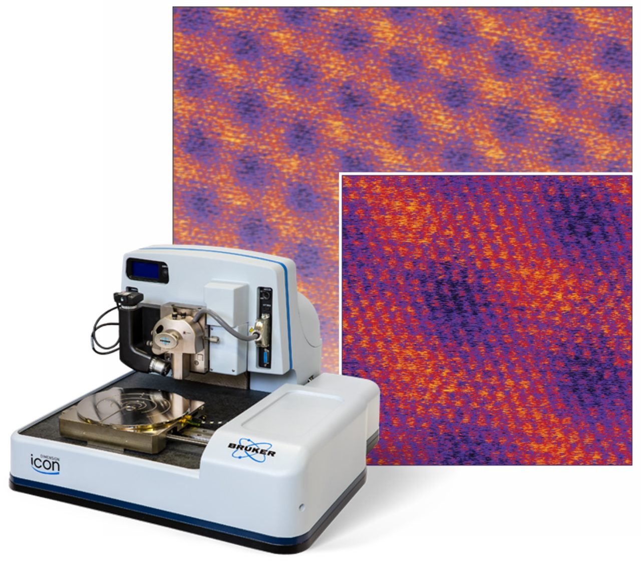
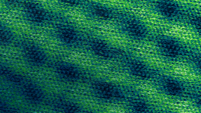
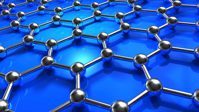
Atomic Lattice Imaging of 2D Materials
Atomic lattice imaging is a key AFM resolution benchmark. Achieving this level of resolution has typically required highly experienced users and mechanically compact AFM systems. With the latter often being at the cost of limited scan ranges and restrictive sample sizes.
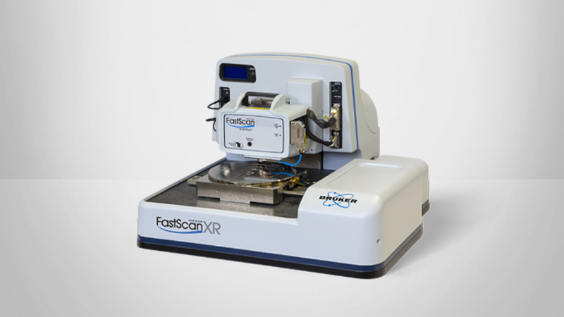
The superior signal-to-noise and ease of operation of TR-DFM, together with the Dimension Icon’s proven highest performance, delivers routine atomic lattice imaging of 2D materials on a large-sample, open access platform.
TR-DFM is a powerful mode for studying 2D materials. It has allowed us to directly visualize both atomic crystal lattices and moiré superlattices formed in vdW materials at unprecedented success rates. The advantages of this mode could have a transformative impact on fundamental and applied research on vdW materials and devices.

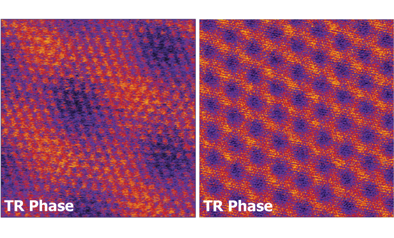
Simplified Nanoscale Characterization of Moiré Superlattices
When 2D materials are assembled into vertically stacked van der Waals (vdW) heterostructures, the interlayer twist creates a moiré superlattice whose period is a function of twist angle. Slight changes in the twist angle can dramatically change the electronic properties of these stacked structures, making them attractive for use in electronic devices, energy storage, and quantum technologies. As such, the ability to characterize vdW heterostructures at the nanoscale is extremely important.
TR-DFM has successfully resolved key structural properties that are known to directly affect the electronic properties of vdW heterostructures, such as:
- Surface and sub-surface moiré patterns for determination of twist angle, strain, and other nanoscale spatial variations
- Atomic crystal lattice structure of individual layer materials for identification of vdW flake orientation
TR-DFM offers key advantages over other AFM modes for straightforward and reliable characterization of moiré superlattices, including:
- Consistent high-resolution imaging
- Compatibility with nonconductive samples and substrates
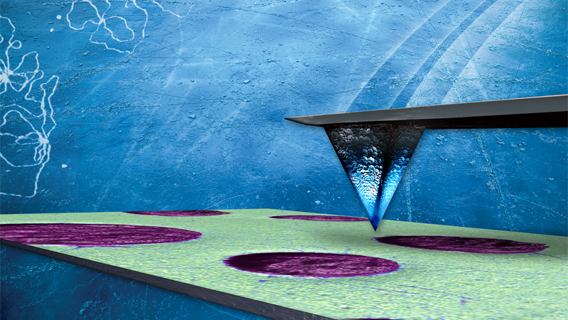
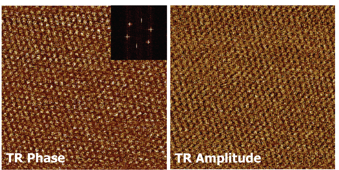
Compositional Mapping of Polymers with Enhanced Sensitivity
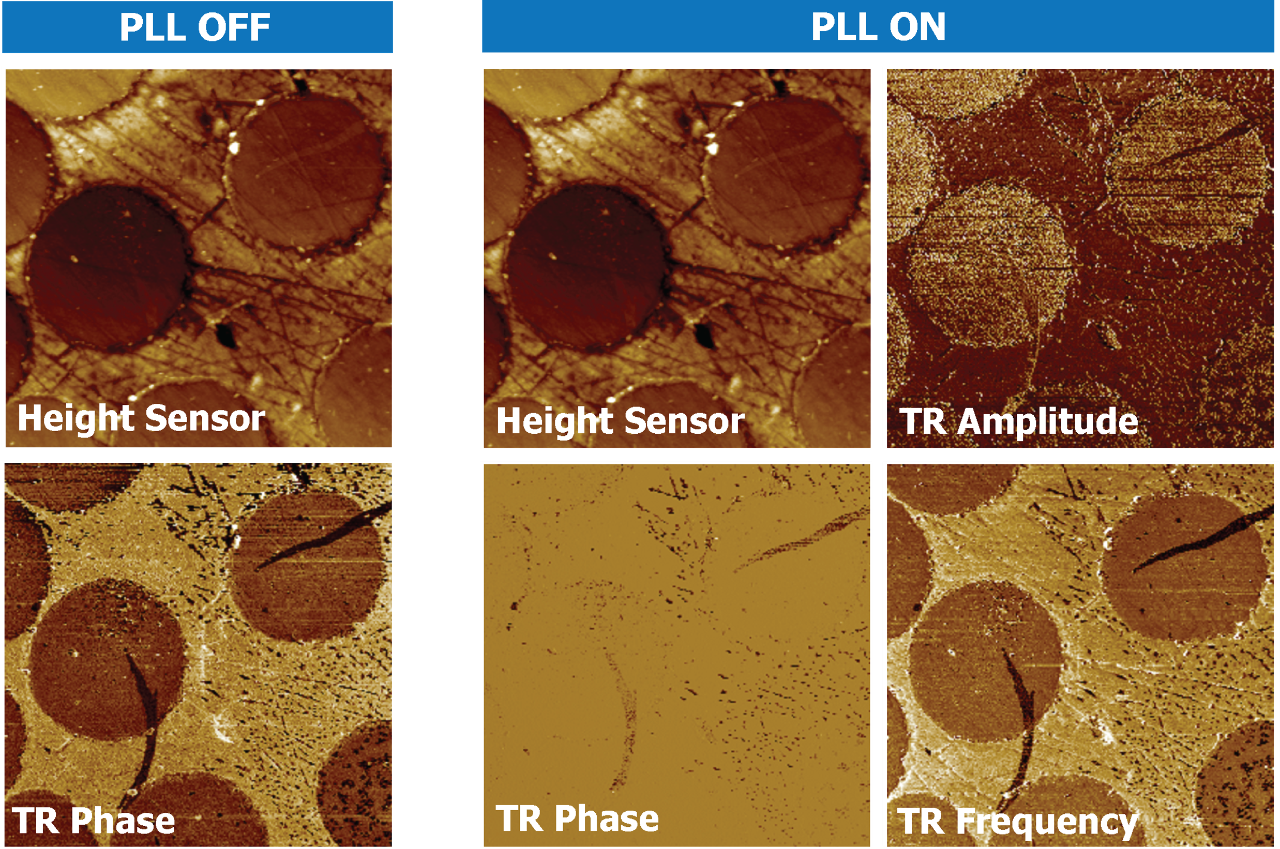
In TR-DFM, the torsional resonance modulation gives rise to strong contrast in the TR Phase image that is related to the mechanical properties of the sample. As a new feature of the NanoScope 6 controller, a Phase Locked Loop (PLL) is now available in TR-DFM. When applied, the PLL adjusts (nulls) the Phase shift and tracks the associated shifts in Resonance Frequency and Amplitude, where contrast observed in the Resonance Frequency image is related to elastic modulus and contrast in the Amplitude image indicates dissipative areas.
Using this PLL in TR-DFM can provide novel information with regards to nanomechanical properties of soft materials, such as polymer blends and composites, by enabling compositional mapping with enhanced sensitivity and insights into dissipation.
Built on the benefits of Torsional Resonance Mode (TR-Mode)
Torsional Resonance mode (TR-Mode) was developed by Bruker in 2003. In TR-Mode, the torsional resonance amplitude (or phase) of the AFM probe is used to control the feedback loop and maintain the tip-surface relative position through lateral interaction. The nature of tip-surface interaction facilitates phase measurements to resolve the in-plane anisotropy of materials, as well as measurements of dynamic friction at the nanoscale.
TR-Mode was first introduced as an alternative to TappingMode for high-resolution topography imaging of soft materials. As tip-sample interactions remain in the attractive regime, TR-Mode has proven to be beneficial for imaging samples with high-adhesion. And because the Q-factor of the torsional resonance of the AFM probe is significantly higher than the flexural resonance Q-factor, TR-Mode has provided enhanced structural information for samples requiring highly sensitive phase detection.
The benefits of TR-Mode have been integrated with other Bruker modes, including:
- Torsional Resonance Dynamic Friction Microscopy (TR-DFM)
- Torsional Resonance Tunneling AFM (TR-TUNA)
- Torsional Resonance Magnetic Force Microscopy (TR-MFM)
- Torsional Resonance Tunneling AFM (TR-EFM)