Single Molecule Force Spectroscopy
Single Molecule Unfolding
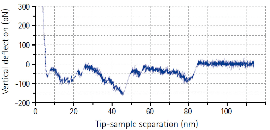
Fibronectin Unfolding
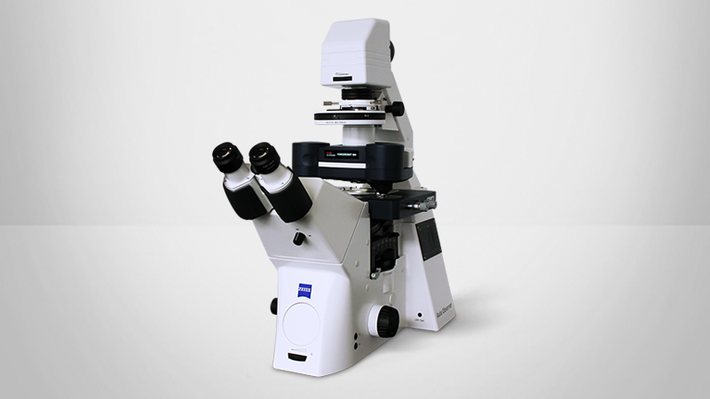
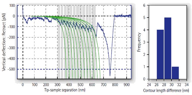
Unfolding curve of native fibronectin, a large modular extracellular matrix protein, imaged with the NanoWizard BioScience AFM. The highlighted fit regions show the successive unfolding of 12 FNIII domains, with WLC fits to each event.
There are 18 domains in a single fibronectin molecule, each has 90 amino acids, and therefore a contour length of around 30nm. The histogram shows a clear peak at 30 nm.
Membrane Protein Unfolding
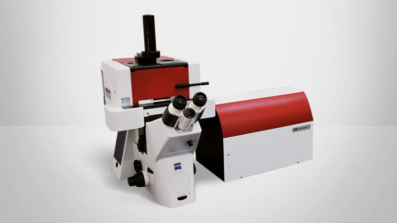
Force spectroscopy curve of a membrane protein unfolding, imaged with the ForceRobot AFM. The structure and kinetics of the G protein-coupled receptor (human β2 adrenergic receptor β2AR) were investigated for a variety of ligands.
Data courtesy of:
Zocher, Fung, Kobilka and Müller, Structure 20, 1391-1402, 2012
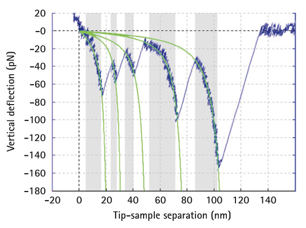
ddFLN Filamin Unfolding
Force spectroscopy curve of ddFLN filamin unfolding, with WLC fits marked, imaged with the ForceRobot AFM. Multiple events are automatically recognized.
Sample courtesy of:
M.Rief, TU München

Bacteriorhodopsin Unfolding
A single Bacteriorhodopsin molecule is unfolded and pulled out of purple membrane, imaged with the ForceRobot AFM.
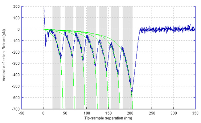
Titin force extension profile
Force extension profile for titin with worm-like chain fits to the unfolding events, imaged with the ForceRobot AFM.
Recognition Maps
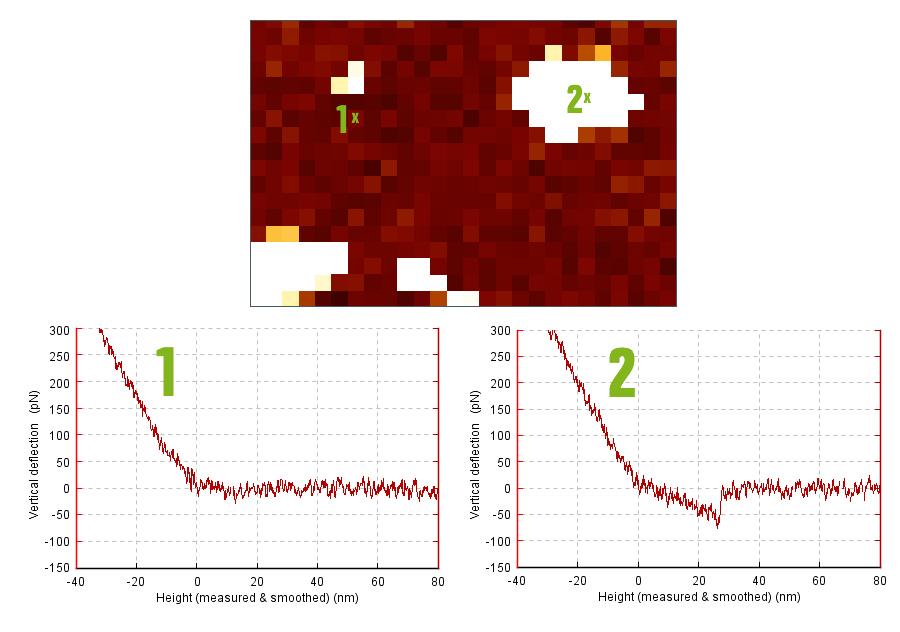
Protein Covered Surface
Example of a recognition map on a protein-covered surface, imaged with the ForceRobot AFM. Recognition events can be clearly identified:
- Top: Recognition Map
- Bottom left: No recognition event (force curve at position 1)
- Bottom right: Recognition event (force curve at position 2)
Single Molecule Manipulation
DNA Pulling
Manipulation of a single DNA molecule using the two optical traps provided by Bruker's NanoTracker optical tweezers platform. The presence of the invisible molecule can be seen from the extension of the left particle from its trap (indicated by the crosshair).
At the same time, the NanoTracker allows the precise and calibrated recording of the force exerted to assess the elastic response of the DNA at increasing tension. The video is sped up 10x.
Fluorescent DNA Manipulation
Manipulation of DNA using Bruker's NanoTracker optical tweezers platform, simultaneous to sensitive fluorescence microscopy. A native (non-fluorescent) DNA molecule is suspended between two optically trapped beads. A continuous fluid flow is applied using the Multichannel LaminarFlowCell. The buffers flowing by can be changed by moving the microscope sample stage between various laminar channels. In the video, the trap-held DNA molecule is suspended successively in a plain buffer and in a flow of fluorescently labeled DNA molecules, coated with fluorescent proteins that specifically bind to the DNA. Towards the end of the video, one such fluorescent DNA molecule connects to the invisible one, like a rope on the washing line.
Such assays can be used to combine quantitative mechanical manipulation using optical traps with protein binding kinetics studies using fluorescence. The combination of techniques makes such assays particularly powerful.
Fluorescent Rad51 proteins courtesy of:
Dr. M. Modesti, CNRS Marseille