Tumor Biology
Providing key insights into tumor morphology, progression and biomarker expression.
Imaging techniques have provided key insights into tumor morphology, progression and biomarker expression. This in turn has enabled earlier detection of tumors and the development of novel treatment strategies, whereby saving lives.
In vivo imaging methods, such as PET and MRI, provide sensitive, cost efficient and high-throughput technique for the longitudinal study of tumor models. Many cancers are associated with a high metabolic turnover and PET using the injection of a radiolabeled glucose analogue can be used to quantify the glucose uptake and provide an indication of how aggressive a tumor is. Similarly, this imaging method has proved particularly beneficial in the detection of molecular markers. Combining the metabolic data of PET with the high-resolution anatomical and functional information provided by MRI further improves the analytic capacity.
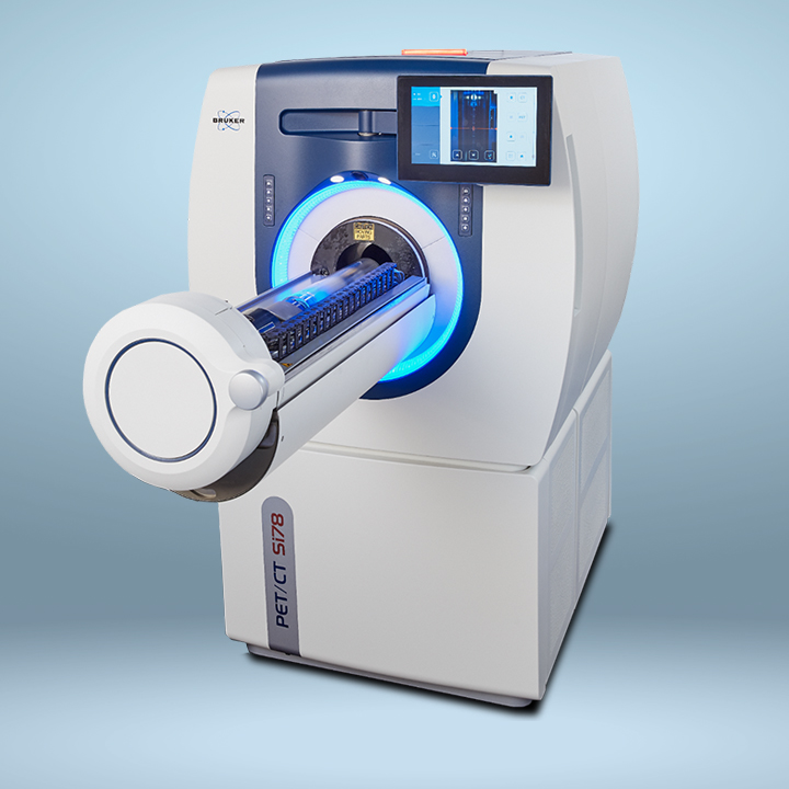
Bruker offers a full range of preclinical multimodal imaging solutions suited to the study of animal tumor models. The Albira preclinical tri-modal PET/SPECT/CT system as well as the the PET/MR 3T combine multiple modalities in a single, easy-to-use instrument to enable whole-body small rodent scans for the localization and quantification of multi-generational metabolic products and biomarkers
Bruker offers a full range of preclinical multimodal imaging solutions suited to the study of animal tumor models. The Albira preclinical tri-modal PET/SPECT/CT system as well as the the PET/MR 3T combine multiple modalities in a single, easy-to-use instrument to enable whole-body small rodent scans for the localization and quantification of multi-generational metabolic products and biomarkers