
MALDI Imaging at the Frontier: Spatial Metabolomics, Single-Cell Analysis, and 3D Exploration in Mixed Reality
Overview
Most of what happens inside a tissue, a tumor, or a 3D cell model is chemically invisible to conventional analysis. The molecular organization is there. The spatial relationships are there. MALDI Imaging is now the tool that makes them visible, from individual cells to complex tissue architectures. This webinar explores what that visibility looks like at the frontier.
In this webinar, Prof. Hopf (CeMOS, TH Mannheim) will present his group's latest findings across the full range of MALDI Imaging-based spatial analysis, from established tissue workflows to single-cell imaging and complex biological systems.
Starting with well‑established tissue workflows, the talk will demonstrate how spatial molecular analysis can uncover altered metabolic and proteomic phenotypes linked to differentiation, disease, or chemical exposure. A particular focus will be placed on toxicology‑relevant applications, showing how label‑free MALDI Imaging enables the unambiguous molecular identification of protein accumulations in kidney tissue, supporting reliable decision‑making in preclinical safety assessment where conventional histology and immunohistochemistry reach their limits.
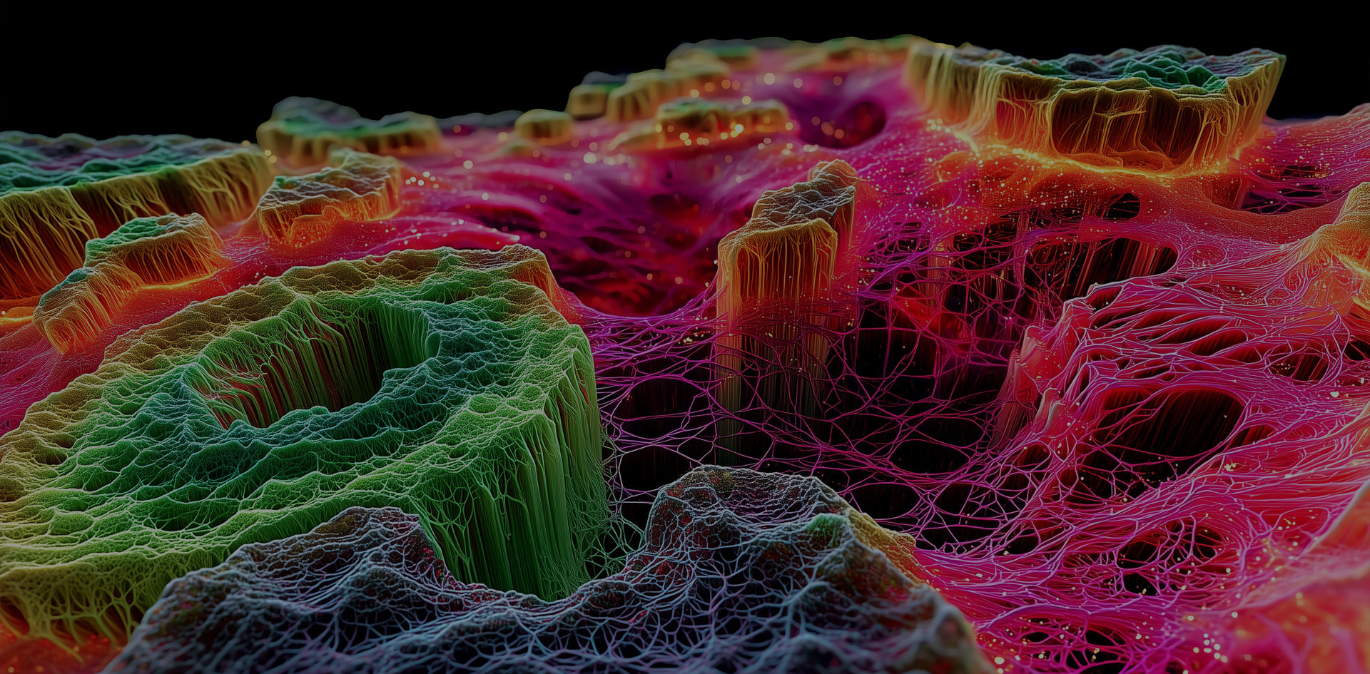
The webinar will then focus on two rapidly developing fields: single‑cell molecular imaging for dissecting cellular activation states, and 3D biology. 3D data. 3D visualization. All in one workflow. Hopf's group applies MALDI Imaging to 3D cell culture disease models including organoids and spheroids, generating volumetric spatial molecular data that can be explored end-to-end in mixed reality, from sample to insight, the geometry is preserved throughout.
Finally, advanced data reconstruction and visualization strategies will be presented that enable researchers not only to analyze but to interactively explore spatial molecular landscapes in mixed reality, opening new perspectives for spatial biology, toxicological research, and translational and pharmaceutical applications.
Key Learning Objectives
- See how MALDI Imaging extends spatial molecular analysis beyond conventional tissue sections into single cells and complex biological systems
- Learn how molecular imaging supports toxicological decision‑making through label‑free, in situ protein identification
- Experience how mixed reality visualization transforms the way researchers interact with complex spatial molecular datasets
- Gain insight into advanced workflows and visualization strategies for complex spatial datasets
- Discover how MALDI Imaging contributes to biological, pharmaceutical, and safety‑related research
Who Should Attend
- Researchers in spatial biology and imaging mass spectrometry
- Scientists working in metabolomics, lipidomics, proteomics, and tissue analysis
- Toxicologists and pharmaceutical researchers involved in preclinical safety assessment
- Researchers interested in single-cell and 3D spatial omics approaches
- Drug discovery scientists using 3D tissue models or organoids for target identification or compound profiling
- Analytical and translational scientists working on biologics, ADCs, or LNP spatial distribution characterization
Speaker

Carsten Hopf, Ph.D., Professor and Head, Center for Mass Spectrometry and Optical Spectroscopy (CeMOS), Technische Hochschule Mannheim, Germany
Prof. Hopf's group at CeMOS has contributed foundational methods in MALDI-based spatial proteomics, metabolomics, and tissue model imaging, with applications ranging from neuroscience to toxicological safety assessment. His group's 3D MALDI Imaging platform was recently published in Advanced Science (2026).
Watch On Demand
Please fill out the form to gain access to the webinar recording.
Nur für die Forschung. Nicht für den Einsatz in klinischen diagnostischen Verfahren.