Deep, Fast, and Large Scale Imaging
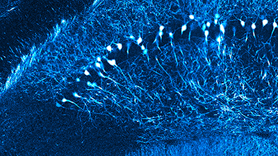
Brain’s electrical activity encodes our experiences, memories, and behavior. Development of two-photon microscopy combined with fluorescent calcium probes made possible for researchers to see the brain in action at high spatio-temporal resolution (1,2). Intriguingly, now researchers can follow activity of large number of neurons and their miniscule processes simultaneously in animal presented with sensory stimulation and engage in behavior.
Bruker offers several developments to allow acquisition and performance of functional measurements under in vivo and in vitro conditions.
Fast acquisition of fluorescent signals coming from calcium probes is enabled with a resonant scanner (3). A superb collection of photons coming from dim samples is guaranteed with sensitive GaAsP detectors located in proximity to a sample. Alternatively, a fibered detection module can be provided to enhance signal collection further. To enable collection of scattered emission photons Bruker’s scopes are supplied with large 2-inch optics. Bruker’s newest scope Ultima 2PPlus offers field of view (FOV) which is over 50% larger in comparison to other manufacturers. With larger field of view simultaneous imaging and photo-stimulation of distant regions in a sample is a dream come through for many researchers (5).
Fast acquisition in 2D is additionally supported with fast volumetric acquisition. This approach is ensured with exclusive piezo devices to drive imaging lens at higher speeds in Z-axis (4). Alternatively, Bruker offers a focusing module which is custom-designed and optically corrected electro-tunable lens (ETL). Positioning ETL within an imaging light path allows for independent Z-positioning of the imaging plane relative to photo-stimulation plane (5).
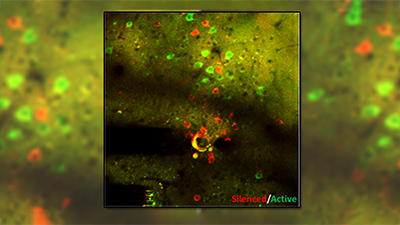
Many of Bruker’s customer implement grin lens, cannula, or prisms to access brain regions deeper than allowed by two-photon microscopy. They successfully perform both imaging and optogenetics experiments with this arrangement (6). Attractively, Bruker’s customers also are supported in applications that require co-registration of signals between miniscope and multiphoton microscopes (7).
Bruker support this application with proprietary software.
References
- 1. Zipfel WR, Williams RM, Webb WW. Nonlinear magic: multiphoton microscopy in the biosciences. Nat Biotechnol. 2003 Nov;21(11):1369-77. doi: 10.1038/nbt899.
- 2. Grienberger C, Konnerth A. Imaging calcium in neurons. Neuron. 2012 Mar 8;73(5):862-85. doi: 10.1016/j.neuron.2012.02.011.
- 3. Turi GF, Li WK, Chavlis S, Pandi I, O'Hare J, Priestley JB, Grosmark AD, Liao Z, Ladow M, Zhang JF, Zemelman BV, Poirazi P, Losonczy A. Vasoactive Intestinal Polypeptide-Expressing Interneurons in the Hippocampus Support Goal-Oriented Spatial Learning. Neuron. 2019 Mar 20;101(6):1150-1165.e8. doi: 10.1016/j.neuron.2019.01.009.
- 4. Goard MJ, Pho GN, Woodson J, Sur M. Distinct roles of visual, parietal, and frontal motor cortices in memory-guided sensorimotor decisions. Elife. 2016 Aug 4;5: e13764. doi: 10.7554/eLife.13764.
- 5. Russell, L. E., Yang, Z., Tan, P. L., Fişek, M., Packer, A. M., Dalgleish, H. W. P., … Häusser, M. The influence of visual cortex on perception is modulated by behavioural state. BioRxiv. 2019. doi:10.1101/706010.
- 6. Jennings JH, Kim CK, Marshel JH, Raffiee M, Ye L, Quirin S, Pak S, Ramakrishnan C, Deisseroth K. Interacting neural ensembles in orbitofrontal cortex for social and feeding behaviour. Nature. 2019 Jan;565(7741):645-649. doi: 10.1038/s41586-018-0866-8.
- 7. https://analyticalscience.wiley.com/do/10.1002/was.00020120