Bone Imaging
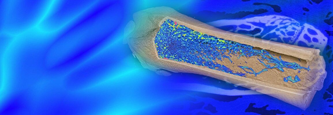
The imaging technique microCT measures absorption of x-rays. This is especially interesting in bones, as the large amounts of calcium and minerals allow you to see bone structure well
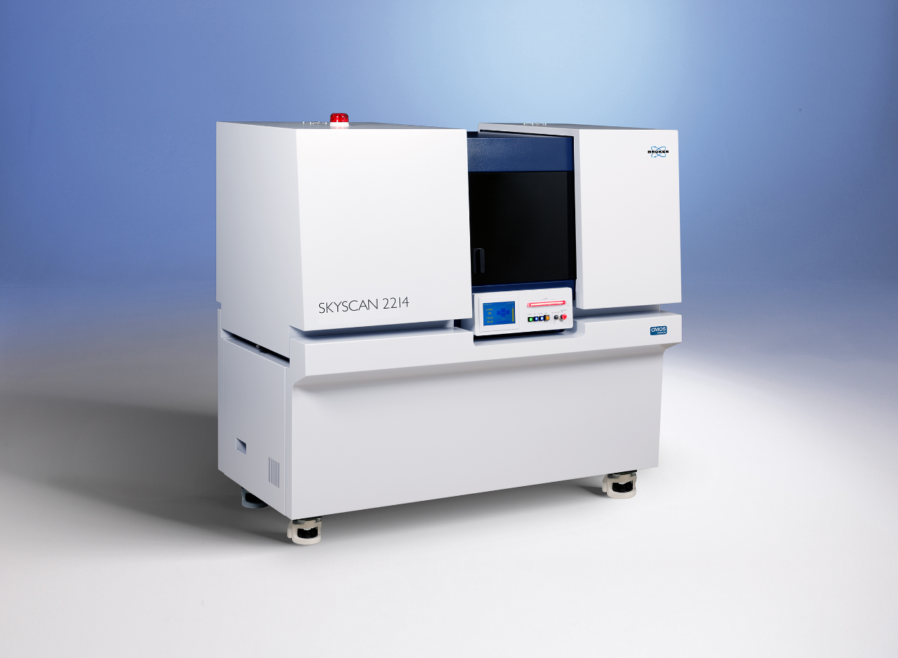
MicroCT is an X-ray attenuation method that provides 3D, high-resolution, non-destructive analysis of the density, architecture and morphometry of hard, mineralized tissues such as bone and teeth.
The most widely used microCT application is bone imaging and the condition most commonly assessed is osteoporosis. However, the scope of this technique is broadening and it is now applied in the study of bone disruption models (fracture, arthritis, tumor), genetic screening or phenotyping, orthopedics, tissue engineering and the evaluation of drug treatments.
Histology remains a widely used technique for bone analysis, as it at offers many advantages such as high resolution and multiple staining options. However, it is time consuming and only offers 2D information. It is also destructive, meaning that in preclinical research, one animal cannot be studied over time. Many animals are therefore required for longitudinal study.
With microCT, these limitations can be overcome. Each sample can be processed in less than one hour to provide detailed, 3D information on bone architecture in a non-destructive way for any site in the animal. In vivo follow up is therefore possible over time, resulting in stronger data, with less animals needing to be used.
MicroCT is applied in the same way as clinical CT. During the scan, radiographs or “projection” images are taken of the sample at multiple angles and then processed using a back-projection algorithm that combines all the information to give a stack of cross-sectional images. These reconstructed images are then processed using a 3D rendering software package that creates 3D models for the measurements of bone morphometry and density.
Bone is the easiest biological tissue to image by X-ray because its calcification means bone structure is easy to visualize, without any sample preparation. Bone imaging will therefore probably always remain the biggest application of microCT in the life sciences.