Solid State NMR Advances Understanding of Osteoporosis
Osteoporosis is the most common metabolic bone disease and our aging population and longer life spans are turning it into a global epidemic. Recent statistics show that one in three women and one in five men over the age of 50 will experience osteoporotic fractures in their lifetime. Currently difficult to diagnose, as there is no clinical manifestation until a fracture occurs, osteoporosis results in a decreased quality of life – as well as a significant burden to healthcare systems across the world. An early diagnosis and assessment of bone structure and density, together with rapid access to treatment, indicates to be the way to prevent this disease - Professor Dr. Sinha and his group approach.
The inherent nature of bone, however, as a solid material, means that it is more difficult to analyse in the laboratory than body fluids or tissue.
A team of researchers at the Centre of Bio-Medical Research (CBMR), Lucknow, India, led by Professor Dr. Sinha is using solid-state nuclear magnetic resonance (NMR) spectroscopy to dive deep into understanding bone and cartilage changes at microscopic levels with the ultimate goal of influencing patient outcomes in osteoporosis.

Professor Dr. Neeraj Sinha
Head of the NMR/MRI unit
Centre of Bio-Medical Research (CBMR)Prof. Sinha is Dean and Head of the Nuclear Magnetic Resonance / Magnetic Resonance Imaging (NMR/MRI) unit at the Centre of Bio-Medical Research (CBMR).
He has worked in the field of NMR spectroscopy for 25 years, starting with his PhD in Physics at the Indian Institute of Science, Bangalore and post doctorate at the University of California, San Diego, where he used solid state NMR to research biomolecules and bioproteins using both theoretical and experimental methods.
The versatility and accuracy of NMR made it the natural choice to advance his work.
Collaboration to advance medical science
The Centre of Bio-Medical Research (CBMR), India, is committed to building a world class research institute. It is dedicated to disease-oriented research and to translating discoveries and observations from the laboratory into new diagnosis and therapies for the benefit of humanity.
Research at the CBMR emphasizes the pursuit of fundamental scientific advancement through interdisciplinary programs and collaborations.
The work of Prof. Sinha’s team focuses on developments in solid state NMR spectroscopy for the study of micro-structural changes in bone due to osteoporosis. Joining the CBMR in 2007, Prof. Sinha continues his pioneering work in the field of biomedical science using Bruker’s NMR spectroscopy as a core part of his research.
The analysis of bone
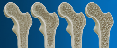
Bone is a composite, amorphous biomaterial consisting of inorganic minerals and organic proteins such as collagen, lipids and water. During osteoporosis, bone becomes susceptible to fracture. Furthermore, recovery following bone fracture in osteoporosis patients is slow. Both factors represent a serious medical condition, particularly in elderly patients.
Solid state NMR spectroscopy is unique in allowing the accurate analysis of bone and other biological systems in their absolute native environments. By also applying dynamic nuclear polarization (DNP)-NMR to the bone samples, new interactions in structural details of bone at sub nano resolution were detected, which help understand the mechanism of osteoporosis disease progression.
The unique impact of NMR
Prof. Sinha’s research group develops and applies advanced solid state NMR spectroscopy to understand various interactions in the bone, investigating components that affect the mechanical properties of bone, particularly in relation to fracture and subsequent healing. There is a current focus on bone hydration and how this can change in the bone as well as in the cartilage due to age, disease and treatment regimens.
The challenge was to develop a research program with the focus on biomedical research into osteoporosis. Using the Bruker 600MHz NMR spectrometer, Prof. Sinha’s team worked closely with the support of Bruker’s applications specialists to design instrumentation and a workflow specifically for bone research. The work set out to measure the differences in bone structure, bone mineral density, and changes over time as the disease progresses.
No time-consuming sample preparation
The nature of bone means that imaging requires specialist instrumentation. Because the water component is bound, there is only a short relaxation time, which means that to quantify bone hydration in its in vivo state, the analysis has to be performed in a short time. NMR offers the major advantage that sample preparation is not required; instead processing in its native state is uniquely enabled by solid state NMR.
Prof. Sinha outlines the benefits: “We are really starting to push the boundaries of bone research, which will be essential to combat the increasing incidence of osteoporosis worldwide. Solid state NMR is the ideal technique as the NMR spectrometer can analyse a small piece of bone to generate analytical spectra. There is no time lost in sample preparation, and the bone can be analysed in vivo to give high-resolution, accurate and meaningful results.”
Investigating bone hydration
To help overcome the challenges of working with bone, the CBMR has worked with Bruker to apply sensitivity enhancement methods including the Bruker BioSolids CryoProbe™, which has improved system performance and the accuracy of results.
The uptake of NMR had traditionally been hampered by its low sensitivity. Now, the higher degree of sensitivity will help to detect rare nuclei such as calcium in the bone matrix to start to map the important role that minerals play in bone density and water content.
Recent research using the BioSolids CryoProbe™ is helping understand the interactions within the native environment of collagen inside bone. The research showed a four-fold sensitivity enhancement in the elucidation of the atomic-level structural details of collagen protein in native state inside the bone.
Water constitutes about a quarter of the cortical bone by volume and can greatly influence the mechanical properties. A key emerging area relating to bone properties lies in differentiating the role of water within the structure. Prof. Sinha and the team designed a series of experiments to investigate how water content affects the bone matrix, a highly dynamic system in which cells undergo continuous regeneration. Using solid state NMR as the spectroscopic method helped elucidate the critical role of water in the bone matrix. Measuring the water-dependent collagen-mineral interface revealed its importance as a parameter to access the bone’s mechanical properties.
Prof. Sinha explains: “Measuring collagen structure, its effect on bone hydration and its association with the interface that underpins the bone structure delivers data that will be used in the design of molecules which bind water content in the bone matrix. The ultimate goals are to support the development of new drugs that help improve patient interventions, and, in the longer term, to develop a clinical diagnostic method based on measuring water content in an in vivo situation.”
Mechanistic insights
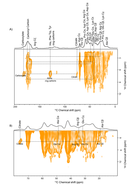
Bone mineralization is a crucial step in bone formation, and collagen present in bones provides a template for the deposition of calcium phosphate (CaP). Investigating interactions between citrate and collagen inside the bone matrix has helped give new structural insights into the biology of bone.
It has long been established that citrate, an abundant structural component of the bone extracellular matrix (ECM), has a high binding affinity to calcium stored in the hard tissue and plays a key role in regulating metabolic functions to maintain the structural integrity of the bone. New research by Prof. Sinha and the team has confirmed that citrate is an integral component of the apatite-collagen nanocomposite in the bone matrix. See Figure 1.
Prof. Sinha adds: “The impressive gain in sensitivity encourages the possibility of performing experiments to elucidate the 3D structural details in the absolute native environment of bones and cartilage at the natural isotopic abundance.”
Shared goals of scientific advancement
“The Bruker contribution to our research has been essential,” says Prof. Sinha. He continues: “Since I joined the CBMR in 2007, Bruker has given us a lot of help with developing specialist instrumentation and workflows that can support the difficulties of working with the bone matrix.”
Future studies at the CBMR will look in more detail at the healing process. Prof. Sinha outlines the next stages of the team’s research: “State of the art equipment such as the BioSolids CryoProbe™ will allow us to build a complete and accurate picture of the changes that the onset of osteoporosis brings as well as the effects of different therapeutic interventions. This advancement applies both to understanding the impact of the disease in the body, and the implications on healing of bone fractures.“
“A deeper understanding of the mechanism of changes that take place as the bone repairs, particularly in the water transfer mechanism, will mean our findings can be used in future drug developments to support and accelerate the bone healing process. We will also continue our investigative work into cartilage, which has a higher water content than bone and is susceptible to osteo-arthritis related degradation.“
“My experience with Bruker has been positive from the start, and I’m very grateful for the expert technical support, to help keep us at the forefront of research in this important field.
We are looking forward to the application of the latest 1.2 GHz high-field NMR systems from Bruker to help advance our research in this critically important field.”
About Bruker Corporation
Bruker is enabling scientists to make breakthrough discoveries and develop new applications that improve the quality of human life. Bruker’s high-performance scientific instruments and high-value analytical and diagnostic solutions enable scientists to explore life and materials at molecular, cellular and microscopic levels. In close cooperation with our customers, Bruker is enabling innovation, improved productivity and customer success in life science molecular research, in applied and pharma applications, in microscopy and nanoanalysis, and in industrial applications, as well as in cell biology, preclinical imaging, clinical phenomics and proteomics research and clinical microbiology.
About the Centre of Bio-Medical Research
The Centre of Bio-Medical Research (CBMR) was established to create a close link among the basic and clinical scientists to fully exploit the potential of magnetic resonance (MR). The important research area of magnetic resonance has produced seven Nobel Laureates in all disciplines of science. Research at the CBMR emphasizes the creation of knowledge in the chemical and biosciences disciplines for application in diagnosis and therapies, the pursuit of fundamental scientific advances through interdisciplinary programs and collaborations, and the education and training of researchers preparing to meet the biomedical challenges of the future.