Understanding Neuronal Activity
Two-fold approach to understand neuronal activation using ultra-high field MRI
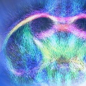
Neurovascular coupling is indirectly measured when blood oxygenation level dependent (BOLD) is used to measure functional magnetic resonance imaging (fMRI). Recent research also suggests that metabolic processes take place simultaneously in the activated cells. To access the role that glucose plays in neuronal activity, researchers from NeuroSpin in Paris and the Weizmann Institute of Science in Israel have explored an alternative metabolic imaging approach based on Chemical Exchange Saturation Transfer (CEST) using ultra-high field (UHF) MRI. The feasibility of utilizing glucoCEST-based fMRI to measure neuronal activity was evaluated in an electrical forepaw stimulation in rats study using a Bruker BioSpec 17.2 Tesla instrument.
The team evaluated the potential of CEST-MRI to record metabolic changes that accompany brain activation. Focusing on glucose, they found a significant change in CEST properties, which they propose is a result of its consumption during neuronal activation.
The negative CEST contrast during stimulation was concurrent with a positive BOLD contrast in the same regions, thus demonstrating the ability of CEST fMRI to locally monitor temporal changes of glucose concentration.
This non-invasive alternative metabolic imaging approach based on CEST-fMRI shows promise for human investigations. It could be expanded beyond glucose detection to investigate additional metabolites that are prominent in neuronal activity.
The Ultra-high Field Advantage
BOLD fMRI greatly benefits from UHF, as the corresponding increased susceptibility effects translate into a greater observable BOLD signal change. Furthermore, the increased spectral dispersion of UHF also benefits CEST imaging, with a high selectivity, high saturation, and a reduction of the exchange rate relative to the chemical shift. Additionally, the SNR that was possible with the BioSpec 17.2 Tesla enabled the approach of this team, something that would have been borderline at low or mid field. This UHF study shows the full potential to drive new discoveries that are not feasible otherwise.
Read the full paper here.
References
Roussel, T., Frydman, L., Le Bihan, D. et al. Brain sugar consumption during neuronal activation detected by CEST functional MRI at ultra-high magnetic fields.
Sci Rep 9, 4423 (2019). https://doi.org/10.1038/s41598-019-40986-9
