Research Highlight:
Katrin Roth, Ph.D., and Lea Wanke
Utilizing Innovative Imaging Technology to Help Solve Complex Research Questions
In this research highlight, Dr. Katrin Roth and Lea Wanke describe their work at the Core Facility Cellular Imaging. At this imaging facility, they help researchers with biological research questions by choosing the best microscopy techniques for their unique samples and imaging requirements. Read the full highlight to learn more about their scientific backgrounds, day-to-day routines at work, and some of the projects they have worked on.

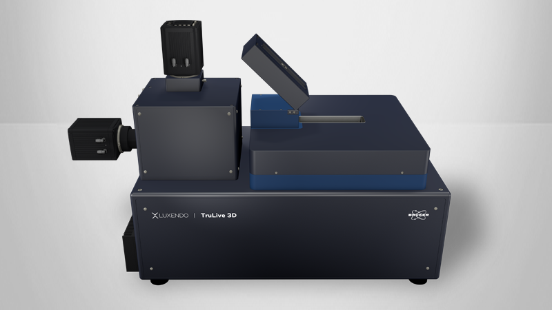
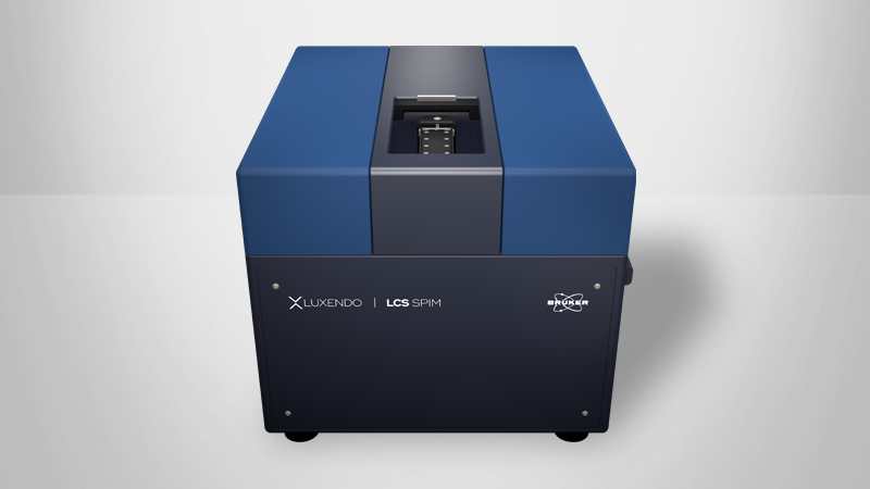
“We have everything from cells to whole animals. With widefield microscopes, we look at fixed cells, either pathological or normal histologically. There are also paraffin-fixed sections and tissue sections. Newer samples include tissue slices, thicker tissue samples, whole organs, and up to full mice.”
Katrin Roth, Ph.D.,
Head of the Core Facility Cellular Imaging

“I like doing microscopy on the large-scale sample microscope with users and teaching them how to image the tissues that they come to the facility with. It’s often organs, like lungs or pancreas, or they take a tissue slice, and then we figure out the best possible way to image them with mounting and clearing techniques.”
Lea Wanke
Scientific Associate
ABOUT THE RESEARCHER:
Katrin is the head of the Core Facility Cellular Imaging at the University of Marburg.
Lea Wanke is a scientific associate at the Core Facility Cellular Imaging at the University of Marburg.
FIELD OF STUDY:
At the Core Facility Cellular Imaging, a range of microscopes are used to examine the movements of cells, cellular interactions, subcellular structures, or even cells within an experimental tumor.
LEARN MORE ABOUT THIS RESEARCH: