Application Note: Long-Term Dual Color Spinal Cord Imaging in Freely Behaving Mice with the nVue LScape Module
Coming from inscopix.com?
You’re in the right place! Inscopix Miniscopes are moving to Bruker.com; in the coming weeks, you’ll see more miniscope details and resources consolidated here.
Same quality, same products, same team – just a smoother, more unified experience.
Image spinal cord activity dynamics in real time
In this application highlight, readers can expect to learn more about the nVue miniscope and LScape module, which allows researchers to conduct imaging of both sides of the spinal cord in freely behaving mice.
Readers can expect to learn more about:
- Recent advancements in long-term spinal cord imaging
- How to perform simultaneous imaging of neural activity using the nVue LScape module
- A novel motion correction method for imaging in awake animals
- How the nVue miniscope and LScape module deliver insights into chronic pain at the cellular level in real time

Introduction
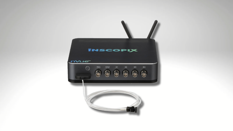
Recent years have seen a rapid proliferation of tools, techniques, and fluorescent sensors designed to characterize neural activity in a naturalistic context in animal models 1. While these advances have permitted refinement of our understanding of circuit dynamics within the brain, investigations further along the neuroaxis have been limited by the technical complexities of surgical preparations to permit optical access to the spinal cord, as well as unique analytical challenges not encountered in conventional neuroimaging. Multiple groups have offered promising solutions to the problem of optical access 2,3. However, local inflammation and fibrosis have previously required re-preparation of the spinal cord for imaging 4, adding layers of risk and difficulty to long-term studies that require stable fields of view and spinal window clarity over multiple imaging sessions. Now, Ahanonu, Crowther, and colleagues have pioneered a novel surgical preparation that makes use of transparent fluoropolymers to inhibit fibrosis within the imaging window 5. Leveraging the wide field of view and high spatial resolution of the nVue LScape module (Figure 1), they performed bilateral, dual color recordings in awake mice during open field arena exploration. Incorporating newly developed motion correction modalities into their analysis pipeline, they tracked neuronal and non-neuronal cells and their responses to nerve injury and noxious stimuli in freely behaving mice with high fidelity over many months, opening the door to previously inaccessible insights into the cellular processes associated with chronic pain states as they unfold in real time.

Capturing spinal cord imaging during free behavior with the nVue LScape Module
Ahanonu*, Crowther*, et al5 outlined a 3-step surgical protocol in either wildtype mice injected with viruses that expressed cell-type specific fluorescent indicators or transgenic reporter mice that permit visualization of glial cells, axons, or cell bodies throughout the CNS or in lamina I dorsal horn projection neurons (SCPNsPhox2a) in the spinal cord. The surgical protocol makes use of easily fabricated, biocompatible materials and specialized fluoropolymers placed at the implant site to inhibit fibrosis and obviate the need for repreparation of the imaging window. Using the nVue LScape module (Figure 2), they uncovered stable responses to noxious and neutral stimuli over time in a naturally behaving mouse, revealing subtle and complex neural dynamics not observed in anesthetized mice. They demonstrated that their preparation does not interfere with normal behavior or locomotion, and introduced a new motion correction method, the large-displacement motion correction method (LD-MCM), to address large rostro- caudal displacement of features of interest along the spinal cord during imaging in awake mice as they respond to stimuli.

The Spinal Chamber
Spinal Chamber
- Stabilizing plates
- Side Bars1
- Designs and CAD files at: https://github.com/basbaumlab/spinal_cord_imaging
- Window Cover
- Spinous Process Needle
- Dental cement, RTV silicone adhesive, and cyanoacrylate adhesive
Surgical Table
- Side Posts
- Clamps
- Arm Rests
- Micromanipulators capable of movement in 3 axes
- Heating pad
- Gas Delivery Scavenger System2
Activity indicator or cell-type marker
- GCaMP expression via AAV delivery
- Wildtype and transgenic reporter mice
General experimental workflow (short protocol) Surgery
A. Viral Injection
- Anesthetize mice and prepare for survival surgery according to institutional protocols.
- Determine optimal virus, volume, titer, and injection parameters for cell types of interest.
- Using a stereotaxic frame and microinjection pump, inject virus retro-orbitally or directly into the spinal cord.
- Suture the skin (for direct injection) and provide supportive post-operative care.
- Wait 1-2 weeks for optimal viral expression.
B. Spinal Chamber and Window
Procedure 1: Install Surgical Window Assembly (2-3 h)
Procedure 2: Laminectomy & Regrowth Inhibition (0.5-1 h)
Procedure 3: Placement of Transparent Optical Window for Long-Term Imaging (0.5-1 h)3.
C. Miniscope Baseplate Installation
- At any time following completion of Procedure 3, attach a Proview Gripper to the stereotax arm or micromanipulator.
- Using the gripper, lower the LScape module, with a baseplate attached, to a few millimeters above the surface of the imaging window. Slowly adjust the coarse focus of the miniscope using the stereotax arm or micromanipulator, then use the e-focus within the IDAS interface to locate the optimal imaging plane.
- Mark the position of the miniscope by zeroing the directional values (AP, ML, and DV) on the stereotax, then apply adhesives (e.g. dental composite and UV-curable glue) to fix the baseplate to the spinal imaging chamber.
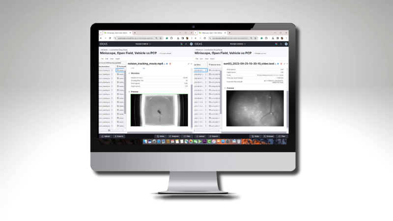
D. Acquire in vivo calcium imaging data with LScape
- Power up the nVue data acquisition system and connect your computer to the system via the DAQ’s internal WiFi or your local area network.
- Ensure that the DAQ has sufficient data storage space and memory for your recording.
- Either anesthetize the animal on a stereotaxic frame or restrain the awake animal* (*the latter is preferred), remove the baseplate cover, and secure the LScape to the baseplate.
- Place the animal in the behavioral arena (for imaging during free behavior) or locate optimal focus in the anesthetized animal and begin recording.
Data Analysis and Results
Processing Behavior and Calcium Imaging Data
After acquisition in IDAS, calcium imaging recordings were processed using CIAtah7,8 and custom MATLAB routines. Registration and motion correction were performed by applying TurboReg10 or LD-MCM to preprocessed movies. LD-MCM was compared to the known motion correction and registration methods TurboReg and NoRMCorre9 and was found to offer superior correction of large rostrocaudal displacement of features of interest (Fig. 3c-d).


Imaging cellular activity during free behavior
Dual color imaging was performed using the nVue LScape module in either restrained or freely moving mice (Fig. 3e-i). Freely moving mice were recorded as they explored an open field arena for up to two hours, during which time they exhibited normal locomotive behavior (Fig. 3j). Bilateral spinal cord imaging data was acquired both from fixed and freely moving animals, a proportion of which had undergone spared nerve injury (SNI), a model of neuropathic pain (Fig. 3k). Behavior data was recorded using multiple high-speed cameras positioned to capture various postural aspects of the mouse’s behavior from different angles. Behavior was analyzed using DeepLabCut6 or the accelerometer in the nVue LScape IMU. Noxious and neutral stimuli were delivered manually while inside a custom chamber (Fig. 3l) or during exploration of an open field arena (Fig. 3m). Fields of view were determined to be stable for over 9 months when recorded with the nVue LSCape module (Fig. 3n-o).
Discussion
Using novel surgical approaches and motion correction workflows, Ahanonu*, Crowther*, et al.5 conducted cellularresolution imaging across a large field of view spanning both sides of the spinal cord in freely behaving mice using the nVue LScape widefield dual color miniscope module. They established a robust preparation for imaging dorsal horn projection neurons that illuminates cellular responses to tissue injury over periods of many months, affording unique new insights into processes associated with chronic pain and neuropathy. Leveraging the large field of view and high spatial resolution of the LScape module, they have begun to disentangle the complex neural responses to both noxious and neutral stimuli in freely moving mice as they naturalistically explored their environments, an experimental paradigm that technical and analytical limitations have precluded until now. They also described a new, deep-learning-based custom motion correction workflow tailored specifically to the large rostro-caudal motion commonly observed in spinal cord imaging. The versatility and modularity of the Inscopix suite of imaging tools also establish a framework for multisite, simultaneous spinal cord and brain imaging, which would grant investigators unprecedented access to the mechanistic underpinnings of the integrated experience of pain and somatosensation along the entire neuroaxis.
References
- Kim, T. H. & Schnitzer, M. J. Fluorescence imaging of large-scale neural ensemble dynamics. Cell 185(1): 9–41 (2022).
- Shekhtmeyster, P. et al. Multiplex translaminar imaging in the spinal cord of behaving mice. Nat Commun 14, 1427 (2023).
- Nelson, N. A. et al. Imaging spinal cord activity in behaving animals. Exp Neurol. 320: 112974 (2019).
- Farrar, M. J. et al. Chronic in vivo imaging in the mouse spinal cord using an implanted chamber. Nature Methods 9, 297-302 (2012).
- Ahanonu*, B., Crowther*, A, et al. Long-term optical imaging of the spinal cord in awake, behaving animals. Nat Methods (2024). https://doi.org/10.1038/s41592-024-02476-3.
- Mathis, A. et al. DeepLabCut: markerless pose estimation of user-defined body parts with deep learning. Nature Neuroscience 21, 1281-1289 (2018).
- Ahanonu, B. & Corder, G. Recording pain-related brain activity in behaving animals using calcium imaging and miniature microscopes. In Seal, R.P. (eds) Contemporary Approaches to the Study of Pain. Neuromethods 178, 217-276 (2022).
- Corder*, G., Ahanonu*, B, et al. An amygdalar neural ensemble that encodes the unpleasantness of pain. Science 363, 276-281 (2019).
- Pnevmatikakis, E. A. & Giovannucci, A. NoRMCorre: an online algorithm for piecewise rigid motion correction of calcium imaging data. J Neurosci Methods 291, 83-94 (2017).
- Thévenaz, P. et al. A pyramid approach to subpixel registration based on intensity. IEEE Trans Image Process 7, 27-41 (1998).
- Dinc, F. et al. Fast, scalable, and statistically robust cellextraction from large-scale neural calcium imaging datasets. bioRxiv 2021.03.24.436279 (2021).