Languages

BioAFM Journal Club

Visualizing Intracellular Nanostructures of Living Cells by Nanoendoscopy-AFM

by Marcos Penedo et al.

Key Points

  • Identified real-time rupture events that correlate to the forces and indentation depths required for the probe to reach different subcellular levels, namely the cytoplasmic and nuclear membranes;
  • Cell viability did not change over the course of 3.5 hours as the researchers repeatedly penetrated the entire cell in order to reconstruct 3D maps of the entire cell; and
  • This technique will allow direct observation, analysis, and manipulation of intracellular and cell surface dynamics and is expected to provide deeper insights into intracellular biological processes.


 Receive future Journal Club updates via email.

This review appeared in the January 2022 edition of the BioAFM Journal Club — a monthly email brief highlighting leading-edge research and the latest discoveries supported by Bruker BioAFM technology.

Sci. Adv. 2021, 7 (52), eabj4990
DOI: 10.1126/sciadv.abj4990

Because AFM is a surface technique, it is usually challenging to perform sub-cellular imaging and study the molecular dynamics of intracellular components. Other factors that contribute to this challenge include the interpretation of complex data (reference force imaging) and decreased cell viability resulting from the high force-loads continuously being exerted on the cells.

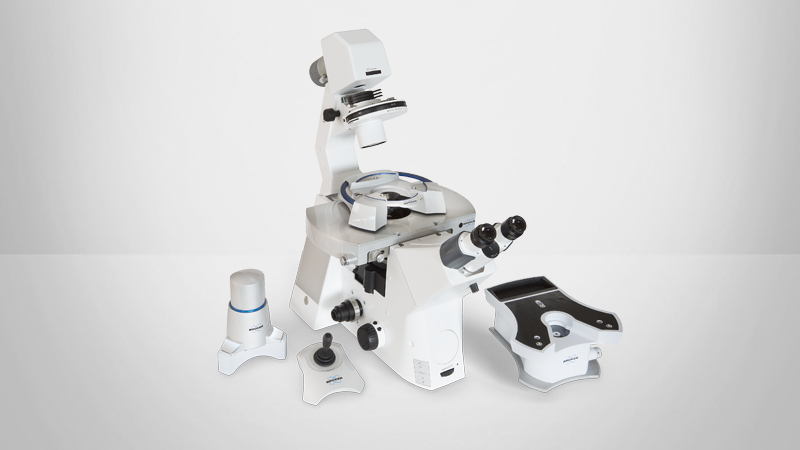
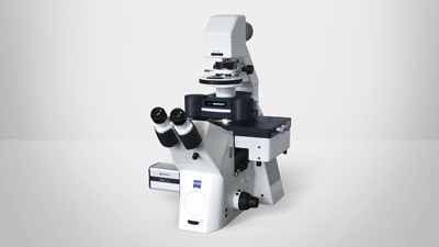
In this article, researchers performed high-resolution AFM and nanomechanical mapping with Bruker's NanoWizard IV BioAFM to study sub-membrane, intracellular structures inside living cells. In the last two decades, AFM has become an indispensable tool for performing high-resolution structural analysis of specimens ranging from single molecules to complex biological systems. The ability of AFM to operate under near-native sample conditions enables the study of a range of dynamic molecular processes that cannot be addressed with other high-resolution tools, such as electron and fluorescence microscopy.

To visualize the interior of the cell, the authors applied needle-like, custom-made nanoprobes long enough to penetrate the cell (3.5 µm) and sharp enough (tip radius < 20 nm, and tip base < 200 nm) to enable high-resolution subcellular imaging. Using force-distance 3D mapping (QI Mode), they identified real-time rupture events that correlate to the forces and indentation depths required for the probe to reach different subcellular levels, namely the cytoplasmic and nuclear membranes. By repeatedly penetrating the entire cell (3D nanoendoscopy) and performing a customized analysis of the rupture event, they were able to reconstruct 3D maps of the entire cell and highlight individual organelles, such as the cell nucleus and cytoskeletal filaments. Cell viability did not change over the course of 3.5 hours, which is most likely because the very small tip base of the needles result in minimal damage to the cell. Furthermore, by verifying the Z-position for different organelles within the cell, the authors were able to perform continuous amplitude modulation (tapping) imaging of the reticular surface of the cytoplasmic side of the cell membrane (2D nanoendoscopy).

This technique will allow direct observation, analysis, and manipulation of intracellular and cell surface dynamics and is expected to provide deeper insights into intracellular biological processes.


      KEY TERMS:

  • Cell Dynamics; Cell Viability; Subcellular Imaging; Tapping AFM