
Bruker Solutions for Organoid Imaging
Complementary Imaging Approaches for Complex Organoid Systems
Imaging organoids can be challenging due to their thickness, cellular heterogeneity, and dynamic behaviors, but complementary techniques now make it possible to meet these demands — providing the ability to label specific cell types, proteins, and pathways and follow how they change during growth, analysis, and maturation. Each technique is optimized for different needs: light‑sheet and multiphoton microscopy enable gentle, long‑term imaging of living organoids, high‑throughput screening microscopy supports quantitative, comparative studies across many conditions, and super‑resolution microscopy reveals nanoscale organization at experimental endpoints.
Understanding organoids and imaging challenges
Organoids are three‑dimensional, stem‑cell‑derived models that can reproduce key structural and functional features of real organs. They enable imaging and analysis of cellular organization, interactions, and processes within a 3D context.
By self‑organizing into tissue‑like architectures, organoids provide researchers with access to human‑relevant biology in controlled, reproducible laboratory systems. They are widely used to study development, disease mechanisms, drug response, and patient‑specific variability.
Organoids capture essential features of real tissues, including cell diversity, spatial organization, and developmental programs, while remaining accessible to controlled experimentation. This makes them powerful models for studying development, disease progression, and therapeutic response using human‑derived cells.
Imaging performance is limited by the three-dimensional structure of organoids, which restricts imaging depth and complicates data acquisition. As samples become thicker and more complex, signal quality can degrade, especially toward the center of the organoid. In live imaging, phototoxicity and photobleaching further limit how long and how frequently samples can be imaged. These constraints require balancing acquisition speed, resolution, imaging depth, and sample impact.
Imaging organoids is more challenging than 2D cultures due to their three-dimensional structure, which limits imaging depth and complicates data acquisition — depth, scattering, and sample heterogeneity all limit image quality. In live organoids, phototoxicity and environmental stability are critical concerns. Imaging strategies must balance penetration depth, resolution, speed, and sample health.
Common Organoid Measurement Needs, by Lifecycle Stage
Organoids evolve over time, from initial self‑organization through experimental manipulation to endpoint analysis. Each stage raises different biological questions and places distinct demands on imaging. Aligning microscopy techniques with these stages ensures that imaging supports, rather than limits, discovery.
Imaging needs shift accordingly: early development emphasizes live, longitudinal observation; analytical phases require higher‑throughput, comparative analysis; and endpoint studies focus on resolving cellular and subcellular structure.
This section is organized around these three stages—development, comparative analysis, and endpoint analysis — highlighting how imaging requirements evolve and how different approaches are used to meet them.
FIND THE RIGHT TECHNOLOGY FOR YOUR MEASUREMENTS:

Organoid Development
Core question(s): How do cells self‑organize into tissue‑like structures? How do cell types emerge, interact, and mature over time?
Importance: Organoid development is dynamic, so capturing growth, morphogenesis, and differentiation requires imaging approaches that can follow intact samples over extended periods without disrupting normal biology.
Technologies commonly used: Light-Sheet Microscopy · High-Throughput Screening Microscopy

Comparative Analysis
Core question(s): How do organoids respond to drugs, genetic perturbations, or environmental changes? Which phenotypic or molecular changes correlate with treatment outcome?
Importance: Comparative analysis experiments often involve systematic comparison across many organoids and conditions. Imaging must be consistent, quantitative, and compatible with live samples over defined time windows.
Technologies commonly used: Multiphoton Microscopy · High-Throughput Screening · Light-Sheet Microscopy

Endpoint Analysis
Core question(s): What nanoscale structural changes underlie observed phenotypes? How are proteins, organelles, and signaling complexes organized within tissue context?
Importance: Fixed endpoint samples allow more aggressive labeling, optical clearing, and sectioning, opening the door to high‑resolution interrogation of cellular and subcellular organization.
Technologies commonly used: Multiphoton Microscopy · Super-Resolution Microscopy
Bruker Technology for Organoid Imaging
Bruker fluorescence microscopy techniques are designed to address the specific challenges of imaging complex, three‑dimensional biological samples such as organoids.
Bruker’s organoid imaging portfolio spans live, longitudinal imaging, high‑throughput screening, volumetric 3D imaging, and super‑resolution analysis. Together, these approaches support the full organoid workflow—from early development and dynamic studies, through treatment and screening, to high‑resolution endpoint characterization. Rather than relying on a single technique, researchers can select from complementary methods optimized for different imaging depths, resolutions, acquisition speeds, and experimental stages.
Use this section to explore our fluorescence microscopy techniques, including core capabilities, typical use cases, and how each approach is applied across organoid imaging workflows.
FIND THE RIGHT TECHNOLOGY FOR YOUR MEASUREMENTS:
| Technique | Organoid development phase | Live vs. Fixed | What it provides | Typically used to assess | Questions commonly answered |
|---|---|---|---|---|---|
| Development | Both | Deep, gentle 3D imaging | Growth dynamics, functional activity | How do cells behave deep within living organoids? | |
| Development, Comparative Analysis | Both | Fast volumetric imaging | Architecture, lineage tracing, responses | How does structure change over time or treatment? | |
| Super-Resolution Microscopy | Endpoint Analysis | Fixed | Nanoscale structural detail | Protein organization, subcellular features | How is molecular architecture altered? |
| Comparative Analysis | Live | Automated, quantitative imaging | Drug response, phenotypic variation | Which conditions produce meaningful changes? |
TECHNOLOGY DETAILS
About Our Multiphoton Microscopes
Best used when you need:
- Live, longitudinal imaging of intact organoids over hours to weeks
- Reliable imaging deep within scattering 3D samples
- Dish‑based workflows that preserve sterility and minimize sample disturbance
What multiphoton microscopy provides:
Multiphoton microscopy uses long‑wavelength excitation to image deep within scattering samples, making it well suited for live organoid imaging where imaging depth and phototoxicity are critical. It enables optical sectioning within thick, three‑dimensional samples while maintaining conditions suitable for longitudinal studies.
Multiphoton microscopy is often used when imaging deeper into organoids is required, or when live imaging must be sustained over time without excessive photodamage. This approach provides:
- Deep optical sectioning with reduced phototoxicity: Supports imaging in sensitive, developing organoids and thicker samples
- Flexible sample configurations: Compatible with organoids in standard culture dishes and multi‑well plates for live and screening workflows
- Adaptable imaging geometries: Enables both inverted, dish‑based experiments and upright imaging approaches on a single platform
Typical organoid questions multiphoton microscopy answers:
- How do cells migrate, differentiate, and function within living organoids over time?
- How do genetic or pharmacological perturbations affect activity deep inside intact 3D tissues?
Typical uses in organoid research:
- Live, longitudinal imaging of organoid development
- Time‑lapse observation of organoid growth and dynamics
LEARN MORE:
About Our Light-Sheet Microscopes
Best used when you need:
- Fast, whole‑organoid imaging at cellular resolution
- Repeated imaging of living samples over hours or days
What light-sheet microscopy provides:
Light‑sheet microscopy illuminates samples with a thin plane of light, enabling rapid volumetric imaging of large organoids while minimizing photobleaching and phototoxicity. It is well suited for imaging whole organoids and dynamic processes over time, where fast acquisition and reduced sample exposure are important.
Light‑sheet microscopy is often used when large 3D datasets are required, or when live imaging must be performed over extended periods with minimal photodamage. This approach provides:
- Efficient acquisition of large 3D datasets: Enables rapid imaging of whole organoids and volumetric structures
- Gentle illumination: Minimizes photobleaching and phototoxicity, making it well suited for fragile 3D cultures
- Flexible sample compatibility: Supports both live and cleared organoid imaging workflows
Typical questions light-sheet microscopy answers:
- How does tissue architecture change during growth, over time, or with treatment?
- How do spatial patterns of cells and signals evolve in 3D?
Typical uses in organoid research:
- Imaging dynamic processes in organoids over time
- Capturing whole‑organoid structures and volumetric datasets
- Time‑lapse imaging of 3D cell culture systems
- Studying organoid development and growth
LEARN MORE:
- READ: E-Book: Light-Sheet Imaging 3D Cell Cultures
- READ: Application Note: Imaging Organoids with Light-Sheet Fluorescence Microscopy
- WATCH: Imaging Breast Cancer Evolution in 3D Organoid Cultures
- READ: Application Note: Light-Sheet Imaging Reveals the Secrets of Neuroscience
- READ: Research Highlight: Katrin Roth, Ph.D., and Lea Wanke
About Our Super-Resolution Microscopes
Best used when you need:
- Nanoscale insight into protein localization and cellular ultrastructure
- High-resolution imaging at experimental endpoints using fixed or sectioned samples
What super-resolution microscopy provides:
Super‑resolution microscopy reveals molecular and subcellular organization beyond the diffraction limit, enabling detailed analysis within complex tissue contexts such as organoids. It is particularly suited for resolving structures and interactions that are not accessible with conventional fluorescence microscopy while preserving spatial relationships within intact samples.
This approach provides:
- Resolution beyond conventional fluorescence methods: Enables visualization of structures not resolvable with confocal or widefield microscopy
- True 3D localization in thick samples: Supports volumetric, single‑molecule localization deep (~50 µm) within organoid structures
- Preservation of spatial context: Maintains tissue‑like organization that is lost in dissociated cell cultures
Typical questions super-resolution microscopy answers:
- How are signaling complexes arranged within tissue?
- What subcellular defects accompany disease‑relevant phenotypes?
Typical uses in organoid research:
- Resolving cell–cell signaling within tissue-like contexts
- Visualizing intracellular trafficking and subcellular organization
- Imaging structures such as cilia and mitochondria
- Studying subcellular features not resolvable with confocal or widefield microscopy
LEARN MORE:
About our High-Throughput Screening Microscope
Best used when you need:
- Quantitative comparison across many experiments or conditions
- Scalable imaging workflows for drug discovery or phenotypic screening
What high-throughput screening microscopy provides:
High‑throughput fluorescence microscopy enables automated imaging and analysis of large numbers of organoids under controlled experimental conditions. It is particularly suited for growth and comparative analysis phases, where comparison across many samples and conditions is required. High‑throughput fluorescence microscopy is often used when population‑level responses and variability must be assessed across multiplexed experiments. Key capabilities include:
- Consistent imaging across samples: Supports standardized acquisition in formats such as 96‑well plates
- Automated acquisition and analysis: Enables efficient, reproducible imaging of large sample sets
- Integration with live and fixed assays: Supports both timelapse imaging and endpoint measurements across screening workflows
Typical questions high-throughput screening microscopy answers:
- Which procedures alter organoid growth, viability, or morphology?
- How variable are responses across organoid populations?
Typical uses in organoid research:
- High‑throughput screening during organoid growth and comparative analysis phases
- Multiplexed analysis across experimental conditions
- Population‑level comparison of organoid responses
LEARN MORE:
Choosing and Combining Techniques for Organoid Imaging
Selecting the appropriate microscopy technique starts with the experimental question and the stage of the organoid workflow. In practice, three factors guide technique selection: the stage of the experiment, the scale of information needed, and the acceptable trade-offs.

What stage of the experiment are you in?
- Development/live imaging: Consider approaches that support long-term, low‑phototoxicity imaging of dynamic processes (e.g., light‑sheet or multiphoton microscopy)
- Growth and analysis: Consider approaches that enable high‑throughput, comparative analysis across conditions (e.g., high‑throughput fluorescence microscopy)
- Endpoint analysis: Consider approaches that maximize spatial resolution and structural detail in fixed samples (e.g., super‑resolution microscopy)

What scale of information is needed?
- Whole‑organoid structure and dynamics: Consider volumetric imaging approaches that capture complete 3D datasets (e.g., light‑sheet microscopy)
- Cellular organization within organoids: Consider techniques that balance resolution and imaging depth (e.g., light‑sheet microscopy)
- Subcellular structure and molecular organization: Consider higher‑resolution techniques that resolve features beyond conventional limits (e.g., super‑resolution microscopy)

What trade-offs are acceptable?
- Long-term live imaging: Prioritize reduced phototoxicity and imaging speed over maximum resolution (e.g., light‑sheet, multiphoton)
- High-throughput comparison: Prioritize consistency, automation, and sample throughput (e.g., high‑throughput microscopy)
- Maximum spatial detail: Prioritize resolution and contrast, typically at the expense of speed and live imaging capability (e.g., super‑resolution microscopy)
What drives imaging requirements and decisions?
Imaging depth becomes a limiting factor when working with thick, dense organoids, where signal quality decreases deeper into the sample. As light penetration is reduced and structures become harder to resolve, especially near the center of the organoid, techniques with improved depth capability are required to maintain image quality and extract meaningful data.
Live imaging enables longitudinal studies of development and dynamic processes, but requires approaches that minimize phototoxicity and support long-term observation. This often limits resolution and imaging speed. In contrast, fixed imaging allows higher-resolution techniques, including those that resolve subcellular structures, but only captures endpoint information. As a result, live imaging prioritizes sample health and temporal information, while fixed imaging prioritizes spatial resolution and molecular detail.
Imaging needs change across the organoid lifecycle, from live development and treatment to fixed, endpoint analysis, requiring different combinations of speed, depth, throughput, and resolution.
Early development and comparative analysis studies often require live, low‑phototoxicity imaging. Endpoint experiments favor higher resolution and molecular detail, often using fixed or sectioned samples. No single technique is optimal for every stage.
Common stage-dependent approaches include:
- Live imaging (light-sheet / multiphoton): Used during early development for live, longitudinal imaging to follow growth and dynamic processes over time
- High-throughput screening microscopy: Used during growth and comparative analysis phases and population-level analysis
- Volumetric (3D) imaging approaches: Applied throughout when complete three-dimensional (3D) volume data is needed to capture whole-organoid structure
- Super-resolution microscopy: Used at endpoint (fixed samples) to resolve subcellular structures not resolvable with confocal or widefield microscopy
As a result, imaging progresses from techniques optimized for live, long-term observation to those focused on throughput and finally to methods that maximize spatial resolution, reflecting that different microscopes are used for different phases of development.
No single technique can capture all relevant information in organoids; different techniques emphasize different scales of biology, from whole‑organoid dynamics to subcellular organization. Using complementary approaches builds a more complete and reliable understanding of organoid behavior.
Common complementary approaches include:
- Light-sheet + multiphoton microscopy: Combined for live, longitudinal imaging, where different strengths in imaging speed, depth, and photodamage support dynamic studies over time
- Live imaging + high-throughput screening microscopy: Used together across development and comparative analysis phases to connect dynamic behavior with population-level responses
- Confocal + super-resolution microscopy: Paired to move from cellular context (~250 nm resolution) to resolving subcellular structures not accessible with conventional methods
- Volumetric (3D) imaging + super-resolution microscopy: Used together to link whole-organoid structure with true 3D localization of subcellular features deep within samples
Using multiple techniques allows researchers to span the full organoid lifecycle, where different microscopes are used for different phases of development, and to balance trade-offs between speed, resolution, depth, and phototoxicity that no single modality can address alone.
Selecting and balancing imaging approaches
Different stages of the organoid lifecycle require different imaging approaches, from live development through comparative analysis to endpoint analysis.
Common stage-specific approaches include:
- Light-sheet / multiphoton microscopy: Used during development for live, longitudinal imaging
- High-throughput fluorescence microscopy: Used during growth and comparative analysis phases for multiplexing and screening
- Super-resolution microscopy: Used at endpoint (fixed samples) to resolve subcellular structures
Live and time‑lapse imaging of organoid development requires techniques that support long-term observation with minimal photodamage and sufficient imaging speed.
Common approaches include:
- Light-sheet microscopy: Rapid 3D imaging with reduced phototoxicity, enabling long-term, live imaging of dynamic processes and whole organoids over time
- Multiphoton microscopy: Deep tissue imaging with reduced out-of-focus excitation, commonly used for live and longitudinal studies in thicker samples
3D imaging is required when organoids must be visualized as intact structures, including internal morphology and spatial relationships across the full volume.
Common volumetric imaging approaches include:
- Light-sheet microscopy: Enables complete three-dimensional (3D) volume data acquisition and imaging of whole organoids from microns to millimeters
- Multiphoton microscopy: Supports volumetric imaging in thicker samples, particularly when deeper penetration is required
Different techniques are required depending on whether the goal is whole-organoid context, cellular detail, or subcellular structure.
Common resolution-driven approaches include:
- Light-sheet microscopy: Provides cellular resolution within whole organoids with large field of view and high-resolution imaging
- Confocal microscopy: Provides optical sectioning with ~250 nm resolution for cellular structures
- Super-resolution microscopy: Resolves subcellular structures (e.g., mitochondria, cilia, trafficking) not accessible with confocal or widefield methods
Organoid imaging requires trade-offs between acquisition speed, imaging depth, resolution, and photodamage depending on the experiment.
Common trade-off strategies include:
- Light-sheet microscopy: Balances speed, depth, and resolution by enabling fast acquisition of large 3D datasets with low phototoxicity
- Multiphoton microscopy: Improves imaging depth and reduces out-of-focus excitation, often at lower acquisition speed
- Super-resolution microscopy: Provides the highest resolution and contrast, but is slower and typically limited to fixed or endpoint imaging
See How Our Instruments Are Being Used
ORGANOID DEVELOPMENT: LIGHT-SHEET MICROSCOPY
Time-lapse imaging of medaka organoid formation

COMPARATIVE ANALYSIS: MULTIPHOTON MICROSCOPY
JF552 labeled neuro-spheroid

COMPARATIVE ANALYSIS: LIGHT-SHEET MICROSCOPY
Lung organoid

COMPARATIVE ANALYSIS: LIGHT-SHEET MICROSCOPY
Tumor organoid

COMPARATIVE ANALYSIS: LIGHT-SHEET MICROSCOPY
Fish-derived organoids differentiating into retinal tissue

COMPARATIVE ANALYSIS: LIGHT-SHEET MICROSCOPY
HeLa cells expressing GFP and mCherry

COMPARATIVE ANALYSIS: LIGHT-SHEET MICROSCOPY
hESC-derived pancreatic spheres

ENDPOINT ANALYSIS: SUPER-RESOLUTION MICROSCOPY
Calbindin-labeled horizontal cells in murine retinal tissue

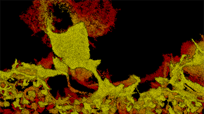
Super-resolution microscopy can be used with organoid models for resolving subcellular structures within tissues.
This image shows calbindin-labeled horizontal cells in murine retinal tissue. Colored by depth from red to yellow across 10 µm. Image courtesy of Nicholas Albrecht and Melanie Samuel, Baylor College of Medicine.
ENDPOINT ANALYSIS: MULTIPHOTON MICROSCOPY
Fusion of two neuro-spheroids

Contact a Bruker Organoid Imaging Expert
Ask us a question, request more information, or get expert instrument and configuration recommendations to fit your specific measurement needs.