Multiphoton Microscopy Applications

Voltage Imaging

Simultaneous recording of membrane potential from multiple neurons with an improved signal-to-noise ratio in vivo

Revealing Circuit Function: Multiphoton Voltage Imaging of Neural Activity

What is voltage imaging?

Voltage imaging is an optical technique that directly measures membrane potential, enabling researchers to record electrical activity such as action potentials and subthreshold dynamics from neurons. Using genetically encoded voltage indicators and multiphoton microscopy, it allows these signals to be captured in intact tissue and even in behaving animals. Recent advances in high-speed imaging have made it increasingly practical to extend these measurements from single cells to neuronal populations, supporting studies of neural circuits and information processing in real time.


Why use voltage imaging?

Understanding how neural circuits function requires measuring electrical activity with both high temporal precision and across many neurons simultaneously. Voltage imaging addresses this need by enabling direct access to the signals that underlie neural communication, supporting studies of fast dynamics at the level of cells and networks.

Voltage imaging delivers:

  • Direct optical readout of membrane potential
  • Millisecond temporal resolution
  • Capture action potentials, subthreshold events, and fast synaptic dynamics

OptoVolt 8x mode (550Hz) showing two-photon imaging of Hippocampal CA1 neurons expressing the voltage indicator FORCE1s, playing at 0.1x speed. Delta F/F in color with anatomical overlay in gray.

Data courtesy of Qiyuan Liang of Michael Häusser’s lab (Hong Kong University), and Francois St-Pierre (Baylor College of Medicine).

Advancing Voltage Imaging Through High-Speed Innovation

The challenge of imaging voltage signals

Despite its potential, voltage imaging has historically been difficult to apply in practice. Fundamental limitations in signal strength and the need for extremely fast acquisition have constrained experiments, making it challenging to scale from single cells to larger neuronal populations.

These limitations stem from:

  • Voltage indicators are dim due to membrane localization
  • Fewer fluorescent molecules cause a low signal
  • Requires kilohertz-scale imaging to resolve spikes


Overcoming barriers to high-speed voltage imaging

Recent advances in multiphoton microscopy are helping to overcome these barriers. As described in recent work, achieving many of the “dream experiments” in voltage imaging requires imaging technology capable of operating at kilohertz speeds while maintaining sufficient signal-to-noise.

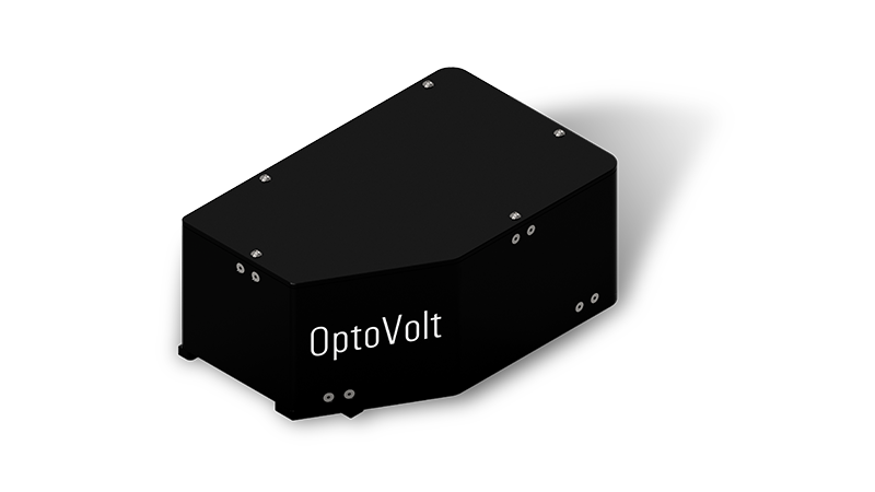
OptoVolt was developed to address these challenges, enabling kilohertz-scale imaging as an add-on to existing multiphoton systems while preserving depth penetration and familiar workflows. By accelerating acquisition and optimizing signal detection, it makes high-speed, population-level voltage imaging more practical in intact tissue.

These advances enable:

  • Kilohertz-scale acquisition
  • Preserves depth penetration and optical sectioning
  • Compatible with established multiphoton workflows

Enabling the Next Generation of Neural Circuit Studies

As voltage indicators and optical instrumentation continue to advance, multiphoton voltage imaging is expected to transition from a specialized technique into a more widely adopted tool for systems neuroscience. Direct optical access to membrane potential enables researchers to observe neural signaling with temporal precision approaching that of electrophysiology, while retaining the spatial coverage and experimental flexibility of optical imaging.

Ongoing improvements in indicator brightness, signal‑to‑noise performance, and high‑speed acquisition are expanding the scale and complexity of experiments that can be performed. These developments are making it possible to move beyond isolated measurements toward population‑level studies of fast electrical dynamics, including action potential propagation, synaptic integration, and circuit‑level computation.

By narrowing the gap between single‑cell electrophysiology and large‑scale optical imaging, multiphoton voltage imaging is opening new opportunities to study how neural circuits communicate, process information, and adapt in real time. As these technologies mature, voltage imaging is poised to play an increasingly central role in uncovering the fast electrical mechanisms that underlie brain function.

Use-Cases

Explore Emerging Applications

A technology that simultaneously records membrane potential from multiple neurons in behaving animals will have a transformative effect on neuroscience research (1,2). Genetically encoded voltage indicators are a promising tool for these purposes; however, these have so far been limited to single-cell recordings with a marginal signal-to-noise ratio in vivo.

The Ultima 2Pplus enabled advanced neuroscience research for Victor Hugo Cornejo's lab in their recent publication, "Voltage compartmentalization in dendritic spines in vivo," where they measured membrane potentials in spines and dendrites in the somatosensory cortex of mice during spontaneous activity and sensory stimulation. To investigate these electrical functions in vivo, they developed a genetically encoded voltage indicator (GEVI) and then used two-photon imaging to measure voltage dynamics. Findings indicate that spines can compartmentalize voltage in physiological states in vivo and has important implications for future studies with synaptic function, synaptic plasticity, and dendritic integration during neurological diseases.

Learn More