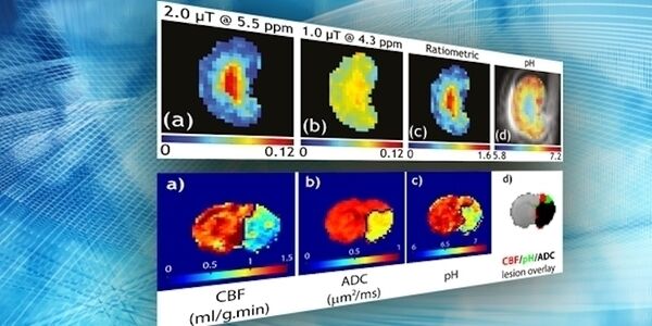

Introduction to CEST for MRI Assessment of Brain Ischemia and Cancer Lesions
Overview
This webinar will introduce the CEST MRI technique and describe its application in small animal imaging. This easily-repeatable, quantitative technique can provide diagnostic information for detecting lesions, deciphering whether they are cancerous, and assessing the effects of treatment approaches as soon as they have started – a capability that would be highly valuable across a range of diseases. The technique is now available in ParaVision 360 and is particularly suited to higher field instruments.
This webinar took place on September 24th, 2020
What to Expect
An introduction to the CEST MRI technique will be followed by a description of its applications in brain ischemia and cancer. A demonstration will be given to show how a basic experiment can be performed and how measuring protein, acid, and metabolite content can be so beneficial. The presentation will be followed by a breif overview of the CEST method introduced in ParaVision 360.
Key Points
- Introduction to CEST MRI for assessing disease parameters in brain cancer and ischemia
- How CEST compliments Bruker’s MRI/PET instruments
- • Assessing tumor metabolism using CEST
- Bruker BioSpin's new CEST method now available in ParaVision 360
- The upcoming CEST 2020 and CEST 2021 meetings, where more can be learned about this product and CEST MRI applications
Who Should Attend?
This webinar will appeal to any researchers and physicians interested in CEST techniques and the 2020 and 2021 CEST meetings. The many applications of this technique will mean anyone in the MRI field, especially physicists, will benefit from learning more about this development.
Dr. Kristin Granlund
MRI Applications Scientist at Bruker USA
Dr. Kristin Granlund is an MRI Applications Scientist at Bruker USA. Prior to joining Bruker, she was a postdoctoral fellow at Memorial Sloan Kettering Cancer Center. Working with Dr. Kayvan Keshari, she developed methods for imaging in vivo cancer metabolism using hyperpolarized pyruvate.
Marty Pagel, PhD
Professor - Cancer Systems Imaging & Imaging Physics MD Anderson Cancer Center
Dr. Marty Pagel has focused on molecular imaging research during the last 20 years in industry and academia. He is a Professor in the Departments of Cancer Systems Imaging and Imaging Physics at the University of Texas MD Anderson Cancer Center. In addition, Dr. Pagel has held leadership positions in professional societies, funding agencies, and scientific journals that focus on molecular imaging. Dr. Pagel’s current research focuses on photoacoustic imaging, CEST MRI, DCE MRI, and PET/MRI, for studies with mouse models of cancer and for clinical trials with cancer patients.