Record DNP Enhancement and Resolution Achieved for Membrane Proteins
Solid-state NMR spectroscopy (ssNMR) represents a promising alternative to X-ray crystallography for proteins and other biomolecules that cannot easily be crystallized in their biologically active forms. Determination of the atomic-level structure of integral membrane proteins, which in many cases require a lipid bilayer to achieve biologically-relevant structure and function, is a promising application of ssNMR that has grown rapidly in recent years.[1] However, to date, membrane protein structures remain underrepresented in the protein data bank, comprising a mere 1% of the nearly 120000 structures currently available.[2] This is especially surprising in light of the variety of diverse functional roles membrane proteins play in biology, from cellular recognition/signaling to channels and transporters. A key reason for the dearth of high-quality membrane protein structures is the difficulty in preparing samples under biologically-relevant conditions and in achieving sufficient NMR sensitivity, or signal-to-noise ratio, on the resulting complex mixtures.
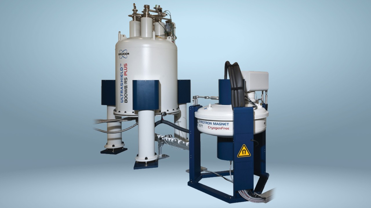
By introducing biradicals into an ssNMR sample and irradiating at the electron paramagnetic resonance (EPR) frequency corresponding to these radicals, dynamic nuclear polarization (DNP) permits the transfer of the much larger electron polarization to the nuclear spins of the sample, thereby vastly improving NMR sensitivity. Since the introduction of DNP, the technique has been able to achieve dramatic enhancements in sensitivity for many types of biological samples. Until recently, however, membrane-embedded peptides and proteins have yielded only modest and highly varied DNP enhancement factors. By applying a direct titration approach to add DNP biradicals to membrane samples in place of conventional centrifugation-based methods, Liao and coworkers were able to achieve significant boosts in DNP enhancement on the transmembrane domain of M2 (M2TM), a proton-selective ion channel in the envelope of influenza A virus. Enhancement factors of approximately 100 and increases in absolute sensitivity relative to room-temperature solid-state NMR of nearly 160-fold have put the DNP performance of membrane samples on par with that of other biological preparations. Further, membrane-embedded peptides were shown to have DNP 13C linewidths as low as 1.0 ppm, among the best reported and only slightly broadened relative to non-DNP values at higher temperature. The study also reports a number of important findings for future membrane-DNP studies:
- The biradical AMUPol outperforms TOTAPOL by a factor of 4, consistent with previous results.
- Glycerol as cryoprotectant provides enhancement factors 1.5-fold higher than DMSO.
- Though enhancement factors may be increased, membranes composed of deuterated lipids do not yield overall higher sensitivity than protonated membranes.
- DLPE lipids yield sharper peptide resonances than DMPC lipids, in disordered peptide regions.
Using paramagnetic relaxation enhancement measurements of the lipid signals, the membrane insertion depths of AMUPol and TOTAPOL were also quantified. While both biradicals show bimodal localization at the membrane surface and approximately 10 Å under the surface, TOTAPOL exhibits a larger membrane-embedded fraction than AMUPol, providing clues toward further sample optimization. With improved enhancement factors and superior resolution, DNP shows great promise for driving rapid advances in membrane protein structural biology.
Full Citation:
Liao, S. Y.; Lee, M.; Wang, T.; Sergeyev, I. V.; Hong, M. Efficient DNP NMR Of Membrane Proteins: Sample Preparation Protocols, Sensitivity, and Radical Location. J Biomol NMR. 2016, 64(3), 223–237.
[1] Hong, M.; Zhang, Y.; Hu, F. (2012) Annu. Rev. Phys. Chem. 63: 1-24
[2] White, S. Membrane proteins of known 3D structure. http://blanco.biomol.uci.edu/mpstruc/; accessed October 24, 2016.