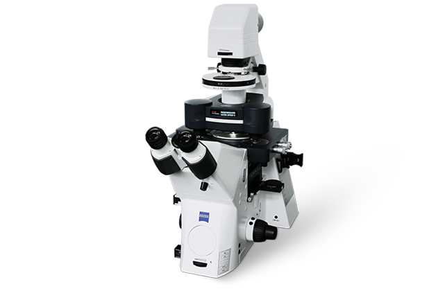
NanoWizard ULTRA Speed 3
Bruker‘s NanoWizard® ULTRA Speed 3 BioAFM sets new standards in speed and performance. Advanced automation features and an unprecedented scanning speed of 1,400 lines per second have been combined in one instrument, that can be seamlessly integrated into advanced optical microscopy and super-resolution techniques. The system delivers correlated sample measurements and the comprehensive nanomechanical characterization of an extensive range of soft matter and living biological samples. Its automated fast-scanning and analysis capabilities maximize throughput and deliver outstanding performance.
Next-Level Performance
NanoWizard ULTRA Speed 3 features a host of new advanced functionalities:
- Fully automated set-up, alignment, and re-adjustment of system parameters, for easy operation and increased productivity
- Bruker’s 3D acceleration sensor and feed-forward technology for unrivalled stability in the tip-scanning motion and highest feedback bandwidth for excellent surface tracking
- DynAsyst for automatic adjustment and optimization of scan parameters in TappingMode™
- Internal LED lighting for stand-alone operation
- QR code reader for improved cantilever handling and management. Scans and automatically sets the parameters of Bruker’s pre-calibrated cantilevers
- Advanced data management and processing capabilities
Morphology: The new software feature DynAsyst and improved Dynamic PID enable easy investigation of fragile, unfixed samples at higher speeds and lowest forces.
Dynamics: A scanning speed of 1,400 lines/sec allows the real-time visualization of dynamic processes.
Nanomechanics: Enables fast, label-free, multiparametric characterization of nanoscale biomechanical properties.
Microrheology: An additional, optional fast Z-scanner with an innovative sensor technology generates reproducible force curves at highest speeds, significantly extending the frequency range for microrheological measurements.
NanoWizard ULTRA Speed 3 sets benchmark standards by delivering fast AFM scanning capabilities that can be combined with advanced optical techniques. Its tip-scanner technology achieves scanning speeds previously unattainable with conventional AFMs.
Empowered Research
Nanoscale structural analysis and nanomechanical characterization deliver crucial insights into functionality at the molecular, single-cell, and cellular levels. NanoWizard ULTRA Speed 3 combines speed, automation, and precision with ease of use. Achieving high-caliber results is easier than ever before and paves the way for a host of new applications and groundbreaking scientific discoveries.
Providing unique advantages
- Stable scanning at speeds of up to 1,400 lines per second
- Seamless integration with advanced optical and super-resolution techniques
- Patented scanner technology
- Latest dynamic PID implementation
- Unparalleled multi-channel data acquisition rates and rapid analysis of complex, multiparametric data sets
Enhancing capabilities
The visualization of dynamic molecular and cellular mechanisms and quantification of the associated kinetics and reaction rates advance our understanding of fundamental biological processes. In conjunction with advanced optics, ULTRA Speed 3 enables multiparametric observation of in-situ dynamics.
- Single-molecule protein dynamics
- Protein folding
- Receptor-ligand interactions
- Mechanosensitive signaling pathways
- Enzymatic reactions
- Kinetics of surface parameters on nanometer scale
Top row images:
Left: Atomic resolution of a calcite crystal plane recorded with TappingMode AFM in water at 60 Hz line rate. 2D FFT analysis (inset) gives rectangular lattice subunit dimensions of a=0.526 nm and b=0.847 nm. Scan size: 19.5 nm × 19.5 nm. Height range: 120 pm
Center: TappingMode in liquid of puC19 DNA using Bruker probe FASTSCAN-D showing majorminor groove resolution (inset). Scan size: 87.5 nm × 87.5 nm. Height range: 3.5 nm
Right: Gasdermin pore-forming proteins imaged in lipid membrane.
Overview: Scan size: 240 nm × 240 nm. Height range: 2.9 nm.
Inset: Scan size: 36 nm × 36 nm. Height range: 3.5 nm. Sample courtesy Han Yu, Daniel J. Müller Lab., ETHZ, Basel, Switzerland
Bottom row images:
Guided thermally driven protein kinetics on DNA nanostructures.
Imaged in TappingMode at 1 frame/sec. Scan size: 150 nm × 150 nm. Height range: 6 nm.
In collaboration with C.M. Domínguez, C.M. Niemeyer, Institute for Biological Interfaces (IBG-1), KIT, Germany.
Next-Generation Automation
Enabling fast results and enhanced productivity
The culmination of Bruker’s pioneering work in the field of BioAFM, NanoWizard ULTRA Speed 3 incorporates cutting-edge innovations and a host of new advanced features. Its high degree of automation, speed, and functionality deliver best-in-class capabilities, significantly increasing the number of samples and positions that can be probed, maximizing throughput and enhancing productivity.
Delivering unprecedented ease of use
ULTRA Speed 3, with an intuitive user interface, customized and favorite workflows, and on-screen context help, is the ideal choice for multi-user environments and imaging facilities. Automated procedures and the choice between standard and advanced features enable experts and less experienced users alike to repeatably produce highest-quality data.
The advanced Data Processing software allows users to browse easily through thousands of images at specific locations, channels, and individual frames in their time-series data, all of which are efficiently saved in HDF5 container format. Various output formats can be exported automatically and simultaneously, such as individual processed data files, image videos or numerical data from cross-sections or histograms.
Taking AFM automation to the next level
- Automated set-up and alignment
- Single-click automated cantilever calibration
- Automated measurement procedures and workflow
- Single-click automated optical image calibration
- Automated scan parameter adjustment with DynAsyst
- Automated, high pixel density mapping and imaging
- Intelligent adaptive scanning routines
- Automated Force Spectroscopy
- Fast, automated scanning of challenging, highly corrugated samples on an inverted microscope
Featuring advanced capabilities
DynAsyst: Automated parameter adjustment in TappingMode for minimal force, utmost consistency, and unattended operation
PeakForce Tapping® with ScanAsyst®: The gold standard for fast and easy high-resolution imaging
PeakForce-QI™: The symbiosis of PeakForce Tapping and QI modes delivers speed and advanced force control for highly delicate samples
ExperimentPlanner: Predefinition of settings and parameters allows complex experiments to run automatically
ExperimentControl: Remote monitoring of long-term lab experiments via a browser on any device
2D DNA origami lattice imaged in buffer with 50 mM NaCl with 1450 × 2048 px and 2.8 μm × 4.0 μm scan size at 20 Hz line rate.
2D DNA nanostructure lattice formation in buffer with 75mM NaCl and 10 mM CaCl2 imaged in TappingMode at 1 frame/sec. Scan size: 1μm × 1μm. Height range: 2.8nm. Sample courtesy of Dr. Adrian Keller, Paderborn University, Germany.
Quantitative Nanomechanics
Enhancing mechanobiology analysis
NanoWizard ULTRA Speed 3 comprises a comprehensive range of tools for the investigation of the nanomechanical properties of samples ranging from single-molecules to single-cells and beyond. An extensive range of modes enable the study of structure, mechanobiology, and dynamics on soft and challenging samples. Perform high-resolution imaging and quantitative nanomechanical characterization of properties, such as Young’s modulus, adhesion, dissipation, and deformation, with optional modes, such as PeakForce-QI, PeakForce QNM®, or QI Advanced.
- Multiparametric characterization in combination with optical microscopy
- Trigger and observe interaction and adhesion processes
- Determine viscoelastic properties
- Contact point imaging (CPI)
- True, real-time force curve monitoring
- Time-dependent, force-induced nanomanipulation
- Easy and precise batch processing of images and force curves
Extending technology
The new NestedScanner microrheology feature extends the operation of multiple Z-piezos, allowing fast analysis of viscoelastic properties. By optimizing use of the Z-piezos, high-speed, high-resolution imaging of steep surface structures with heights of up to 8 μm, e.g., living cells, bacteria, and tight junctions, is now simple.
PeakForce-QNM image of a thin film of styrene-ethylenebutylene triblock copolymer (Kraton G1652) prepared on a silicon wafer. The topography is shown on the left and the corresponding Young’s modulus is shown on the right. Scan size: 1 μm × 1 μm. Height range: 15 nm.
Modulus range: 400 MPa.
Redefining flexibility
The novel SmartMapping feature allows the flexible selection of multiple, user-defined 2D force maps. Numerous regions of interest (ROI) can be selected in advance and examined automatically, enabling the systematic study of large sample areas. The optimal range of force acquisition is continuously evaluated and automatically adjusted by XY and Z-motors in combination with the piezo scanners, delivering a degree of precision and flexibility second to none.
Integration into Advanced Optical Techniques
Setting new standards in correlative microscopy
NanoWizard ULTRA Speed 3 can be seamlessly integrated into advanced optical techniques. Optical guidance allows the fast and easy combination of measurements, while optimizing output and efficiency.
Enabling groundbreaking discoveries
The ability to trigger, control, and observe molecular reactions and cell-cell or cell-surface interactions in real time can significantly improve our understanding of fundamental biological mechanisms and chemical and physical processes.
- One-click optical image import
- Advanced calibration algorithms and visualization routines for precise correlation of AFM and optical data
- Easy navigation around the sample and selection of ROIs in optical image for combined AFM experiments
- SmartMapping for selection of multiple, free-hand drawn ROIs for AFM operation
- Automated large-area, multi-region imaging (by combining DirectTiling, DirectOverlay 2, and MultiScan software features) extends the optical viewing field for atomic force microscopy
NanoWizard ULTRA Speed 3 integrates seamlessly into advanced optics:
- Transmission illumination (brightfield, phase, DIC) using standard condensers and reflection microscopy
- Confocal microscopy and spinning disc
- Ca2+ imaging
- Macroscope combination
- 980 nm optical beam deflection (OBD) option
Optics: Confocal laser scanning fluorescence image of live E.Coli bacteria labelled with Hoechst 33342 (blue - live) and Propidium Iodide (red – dead) acquired on coverslip in buffer.
AFM: NestedScanner TappingMode AFM topography images acquired at 5 Hz line rate (zoom) and 1 Hz line rate (overview)
Overview: Scan size: 4 μm × 4μm. Height range: 1.4μm
Zoom: Scan size: 200 nm × 200nm. Height range: 15nm
- Optical super-resolution (STED, PALM/STORM, FLIM)
- FRET, FCS, FRAP, TIRF, IRM, SIM
- Upright Optics for tissues, implants etc.
- Optical tweezers with OT-AFM
Selected Publications Using the NanoWizard ULTRA Speed Technology
- Xia, M et al., Varying Mechanical Forces Drive Sensory Epithelium Formation. Sci. Adv. 9 (44), eadf2664 (2023).
- Pothineni, B. K et al., Assembly of Hexagonal DNA Origami Lattices on SiO2 Surfaces. Nanoscale 15 (31), 12894–12906 (2023).
- Blaimschein, N. et al., The Insertase YidC Chaperones the Polytopic Membrane Protein MelB Inserting and Folding Simultaneously from Both Termini. Structure 31 (11), 1419-1430.e5 (2023).
- Wang, C. et al., Secreted Endogenous Macrosomes Reduce Aβ Burden and Ameliorate Alzheimer’s Disease. Sci. Adv. , 9 (21), eade0293 (2023).
- Yao, X. et al., Targeted Suppression of Peptide Degradation in Ag‐Based Surface‐Enhanced Raman Spectra by Depletion of Hot Carriers. Small 18 (52), 2205080 (2022).
- Webby, M. N. et al., Lipids Mediate Supramolecular Outer Membrane Protein Assembly in Bacteria. Sci. Adv. 8 (44), eadc9566 (2022).
- Yvanoff, C. et al., Development of Bone Cell Microarrays in Microfluidic Chips for Studying Osteocyte-Osteoblast Communication under Fluid Flow Mechanical Loading. Biofabrication 14 (2), 025014 (2022).
- Konrad, S. F. et a., High-Throughput AFM Analysis Reveals Unwrapping Pathways of H3 and CENP-A Nucleosomes. Nanoscale 13 (10), 5435–5447 (2021).
- Wang, Y. et al., Clustering of Death Receptor for Apoptosis Using Nanoscale Patterns of Peptides. ACS Nano 15 (6), 9614–9626 ( 2021).
- Berger, R. M. L. et al., Nanoscale FasL Organization on DNA Origami to Decipher Apoptosis Signal Activation in Cells. Small 17 (26), 2101678 (2021).
- Fang, T. et al., Spatial Regulation of T-Cell Signaling by Programmed Death-Ligand 1 on Wireframe DNA Origami Flat Sheets. ACS Nano 15 (2), 3441–3452 (2021).
- Scheckenbach, M. et al., DNA Origami Nanorulers and Emerging Reference Structures. APL Materials 8 (11), 110902 (2020).
- Zhao, L.-S. et al., Structural Variability, Coordination and Adaptation of a Native Photosynthetic Machinery. Nat. Plants 6 (7), 869–882 (2020).
- Kol, M. et al., Optical Manipulation of Sphingolipid Biosynthesis Using Photoswitchable Ceramides. eLife 8, e43230 (2019).
- Heesterbeek, D. A. et al., Bacterial Killing by Complement Requires Membrane Attack Complex Formation via Surface‐bound C5 Convertases. EMBO J. 38 (4), e99852 (2019).
Ultimate Versatility with Widest Range of Accessories and Modes
Delivering maximum control
The new Vortis 2.2 controller delivers highspeed, low-noise DACs, and precise position sensor readout technology. Extended channel capabilities support the new proprietary 3D acceleration sensor technology for advanced feed-forward operation, high-performance data processing, and computing algorithms. The latest FPGA technology and dual core PowerPC ensure benchmark data processing rates. The large number of feedback modes and a powerful HV amplifier module facilitate high-speed applications. Passive cooling lowers acoustic noise during the measurement.
Providing flexibility and modularity
NanoWizard ULTRA Speed 3 supports the most extensive range of accessories and modes on the market today, greatly extending the range of applications and experiments possible. Accessories, such as the temperature and environment control options, e.g., gas/fluid exchange modules for perfusion and electrochemistry applications, enable the controlled, systematic study of complex phenomena. The properties of Bruker pre-calibrated cantilevers, manufactured at our fabrication facility, can be integrated into the SPM environment by a single click with the new QR-code reader option, significantly simplifying the initial steps of the experiment.
Comprehensive Environment Control
Temperature control options:
- Ambient to +300 °C with High Temperature Heating Stage (HTHS)
- -35 °C to +120 °C with Heating Cooling Module (HCM)
- -120 °C to +220 °C with CryoStage
Fluid cell options:
- BioCell
- PetriDishHeater (PDH) and PetriDishHolder
- Humidifier for PDH
- CoverslipHolder
- SmallCell for harsh environments
Superior Versatility
- Conductive AFM, EFM, and KPM modules (also with enclosed volumes)
- New Ringing Mode option
- STM module
- Piezoresponse Force Microscopy (PFM) module
- ECCell Electrochemistry cell Scanning electrochemistry (SECM) package Glovebox solutions on request Extensive user management
Enhanced Usability
- HybridStage and motorized stages for automated, large scale sample analysis
- New head elevation kit for samples of additional 8 mm in height
- Head-up stage for samples of up to 121 mm in height
- StretchingStage for following sample property changes under mechanical load
- TopViewOptics for opaque samples
Accessories to extend your BioAFM capabilities
Bruker offers an extensive range of BioAFM system add-ons, accessories, and modes to deliver maximum experimental and sample control, superior versatility, and enhanced useability. These options extend the range of applications and experiments supported by Bruker BioAFMs far beyond what is possible with any other BioAFM system on the market today.
Available options include optical systems/accessories, electrochemistry solutions, electrical sample characterization, environmental control options, software modules, temperature control, acoustic and vibration isolation solutions, and more.
Browse our online accessories database or download our accessories handbook to learn more.
Watch Recent BioAFM Webinars
Our webinars cover best practices, introduce new products, provide quick solutions to tricky questions, and offer ideas for new applications, modes, or techniques.
Testimonials
"The new, automated NanoWizard ULTRA Speed 3 combines high-resolution and high-speed capabilities with excellent ease-of-use. It is ideal for the reliable investigation of single molecule phenomena."
Stefania A. Mari, Daniel J. Müller Lab. ETHZ, Basel, Switzerland
"The ease of integrating the ULTRA Speed 3 AFM with fluorescence microscopy and co-localizing measurements, even on living cells, is unique. Correlating fast multiparametric, nanomechanical data sets with advanced optical images allows cross-validation and delivers novel, reliable results."
Assoc. Prof. Clemens Franz, WPI NanoLSI, Kanazawa University, Japan
"The new NanoWizard ULTRA Speed 3 is an ideal tool for studying mechanobiology in samples ranging from individual molecules to cells and beyond. Its speed, automation, and integration into our optical systems make it perfect for our multi-disciplinary research environment"
Dr. Javier Tamayo, Bionanomechanics Lab., CSIC, IMN, Madrid, Spain

