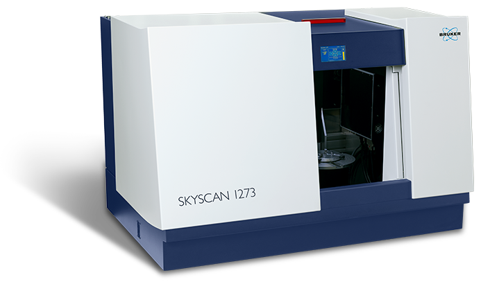
SKYSCAN 1273
A benchtop that fits all
Highlights
SKYSCAN 1273 – High-Capacity XRM
The SKYSCAN 1273 sets a new standard for non-destructive testing (NDT) with benchtop instruments, providing a performance previously only achieved by floor standing systems. Samples with up to 500 mm length, 300 mm diameter, and a maximum weight of 20 kg can be inspected.
The combination of a higher-energy X-ray source running at higher power and a large format flat-panel detector with ultimate sensitivity and readout speed provides excellent image quality in just a few seconds.
SKYSCAN 1273 is complemented by 3D.SUITE. This comprehensive software suite covers GPU-accelerated reconstruction, 2D/ 3D morphological analysis, as well as surface and volume rendering visualization.
Key Features
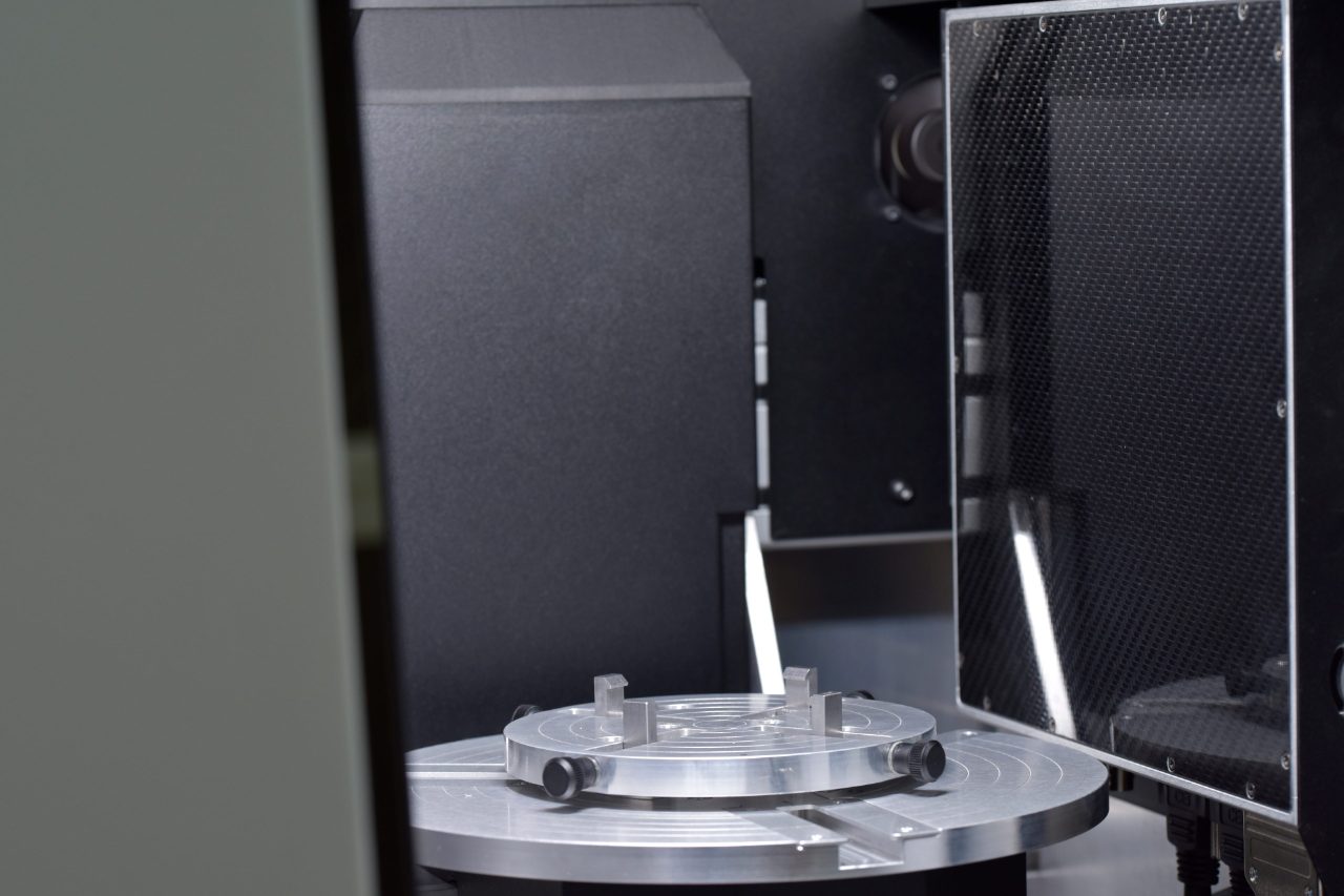
High-capacity benchtop
The SKYSCAN 1273’s large sample chamber accepts samples larger than what can be scanned with a single detector field-of-view. By offset scanning in sections and putting the large format flat-panel detector in offset positions, SKYSCAN 1273 scans large objects up to 250 mm in diameter and 250 mm in length. 3D.SUITE automatically and seamlessly stitches the oversized images together.
Latest detector technology
SKYSCAN 1273 comes equipped with a large-format 6 MP detector based on the latest flat-panel technology, achieving very high contrast in the accumulated images thanks to the large dynamic range. The fast frame rate in combination with an optimized scintillator enable excellent image quality in a stunningly short cycle of less than 15 seconds, which is ideal for time-resolved 3D X-ray microscopy.
In-situ stages
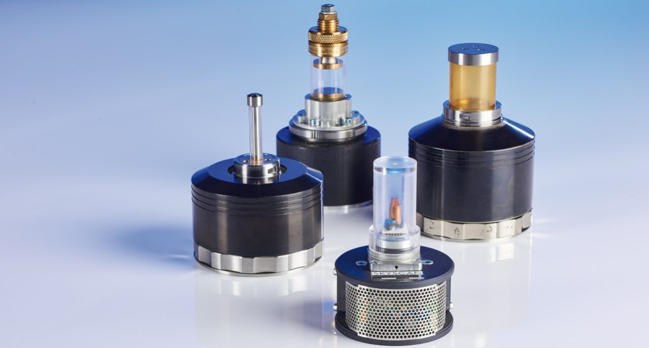
The Bruker material testing stages are designed to perform compression experiments up to 4400 N and tensile experiments up to 440 N. All stages automatically communicate through the system’s rotation stage, without the need of any cable connections. Using the supplied software, scheduled scanning experiments can be set up.
Bruker's heating and cooling stages can reach temperatures of up to 80ºC, or 30ºC below ambient temperature. Just like the other stages, no extra connections are needed, and there is an automatic recognition of the stage. Using the heating & cooling stages, samples can be examined under non-ambient conditions, to evaluate the effect of temperature on the sample’s microstructure.
Highlighted Applications
Bone Applications
The SKYSCAN 1273 builds on Bruker’s key role in the bone research community by offering a microCT scanner for your really big samples. You can put your head inside it’s cavernous sample chamber. So bone biologists and dentists can be joined by dinosaur palaeontologists and archaeologists as well as clinical practitioners imaging biopsies and orthopedic surgery resections, as users of “big microCT”.
- Samples half a meter high and 40cm wide accommodated, scan field of view 25cm diameter and 25cm height – this will accommodate even the largest human and Neanderthal skulls.
- Applied voltage up to 130kV with filters of aluminum, copper and molybdenum give x-ray transmission and optimal contrast over a tremendous range of mineralized biological sample including fossil material mixed with soil and rock.
- Optional mechanical testing and temperature control stages extend the possibilities for biomechanical and orthopedic testing for your clinical or large animal samples.
- Bone morphometry (ASBMR nomenclature) with comprehensive 3D and 2D parameters, with densitometry including BMD calibration references over a 2-32mm diameter size range.
Dentistry
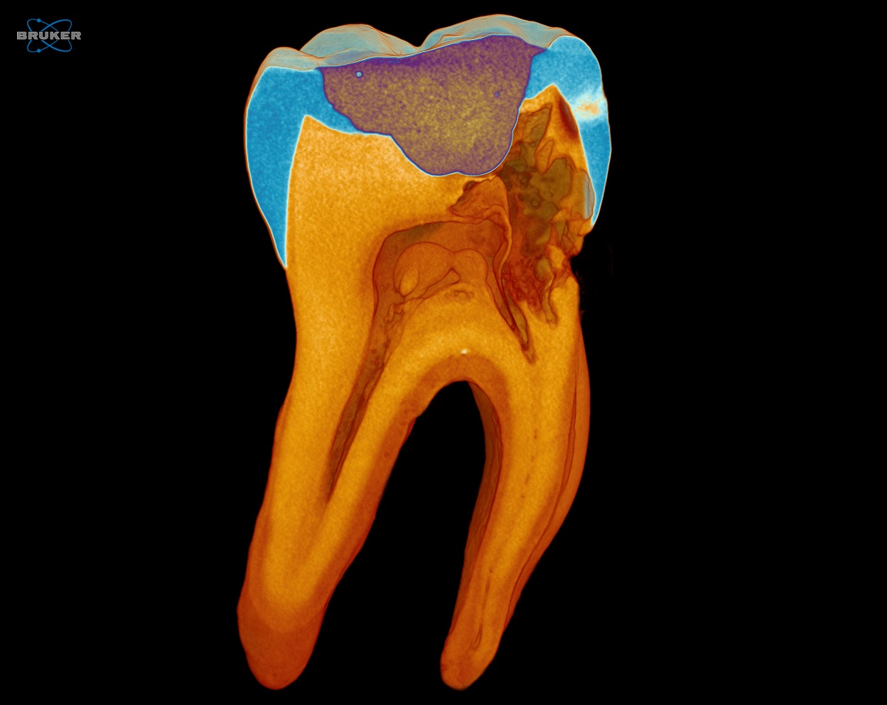
Teeth are a step up from rodent bones in thickness and mineral density, requiring an appropriate microCT solution. The SKYSCAN 1273 is this solution, effortlessly imaging small and large teeth, even fossil ones subject to mineral diagenesis. And this capability in a user-friendly high-energy desktop instrument.
- Fast flat panel with ~3000 x 2000 pixel format, for a huge field of view.
- The large x-ray camera with excellent high energy sensitivity makes most vertebrate teeth scannable as well as prostheses and dental implant materials.
- Bone morphometry (ASBMR nomenclature) with comprehensive 3D and 2D parameters, with densitometry including BMD calibration references over a 2-32mm diameter size range.
Plant and Animal Biology

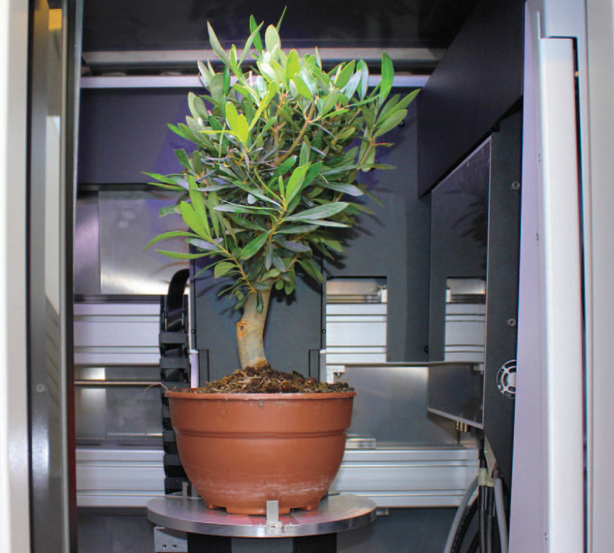
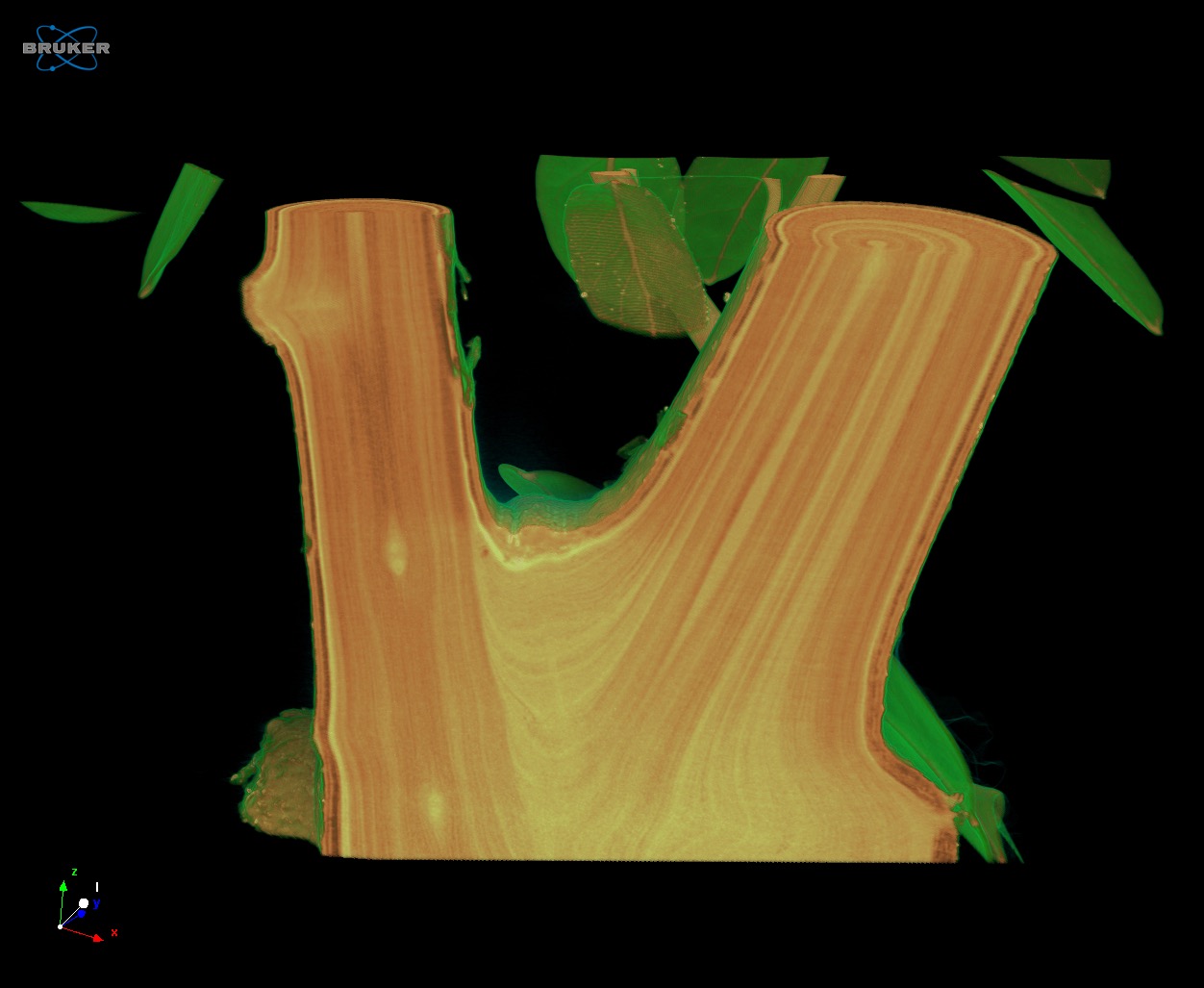
Wood gives exceptionally good results with microCT visualization - the finest details of the xylem and phloem canals are resolved and contrasted. MicroCT differentiation of tissue densities is nondestructive. The enormous scan chamber of the SKYSCAN 1273 allows even small potted trees to be mounted for scanning. A wide range of different plant and animal samples can be visualized and analyzed with little or no need of sample treatment.
- A big scanning chamber for large samples while still a desktop scanner taking up modest workspace makes this the ideal instrument for multi-purpose research.
- Applied voltage up to 130kV with filters of aluminum, copper and molybdenum give x-ray transmission and optimal contrast over a tremendous range of sample types including fossil material mixed with soil and rock.
- Multiple sample scanning possible with high throughput due to the batch-scanning mode, effectively reducing down-time.
- Comprehensive 3D image analysis capability including morphometry and densitometry, 3D registration, segmentation and advanced image processing methods.
Application Gallery
Volume rendered 3D image of a small tree, scanned in the SKYSCAN 1273 without removing from the flowerpot.
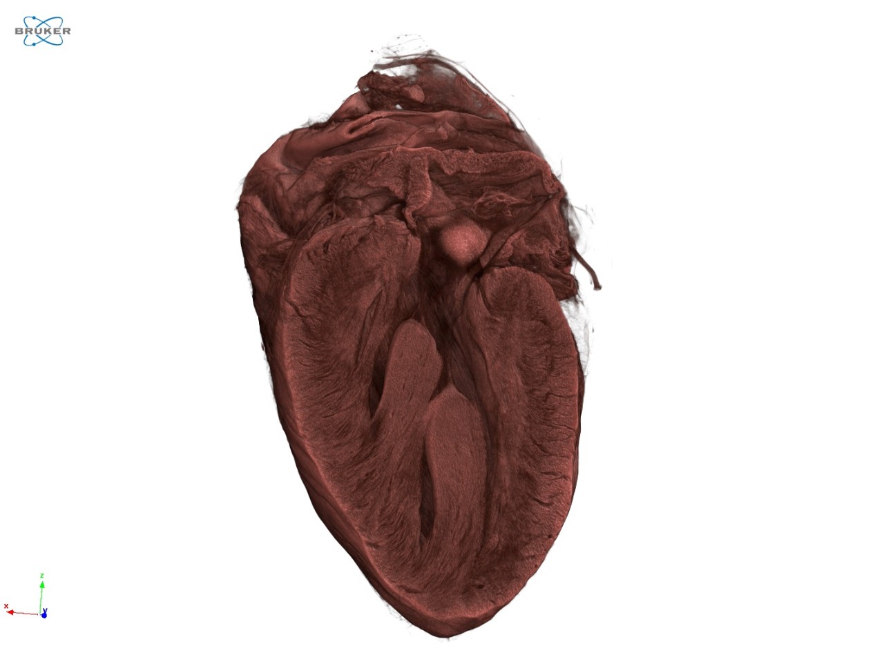
3D rendered image of a stained rabbit heart, scanned at 10 µm voxel size.
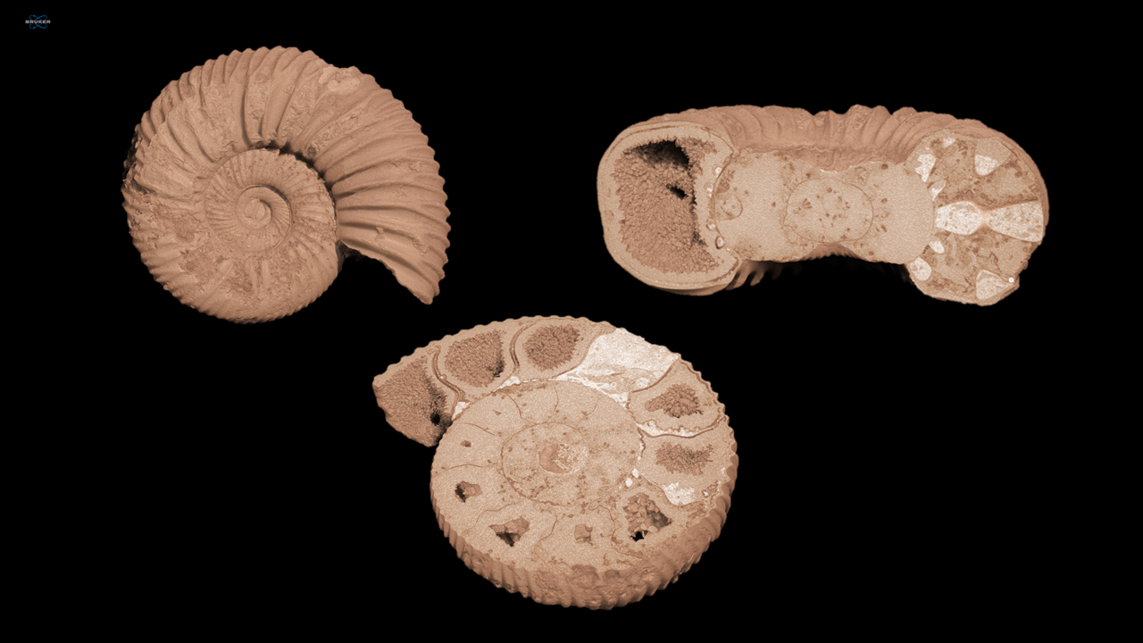
3D illustrations of an ammonite, scanned at 11.5 µm voxel size.
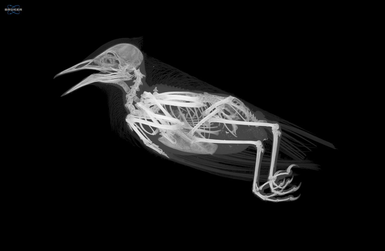
Maximum intensity image of a blackbird, scanned at 30 µm voxel size.
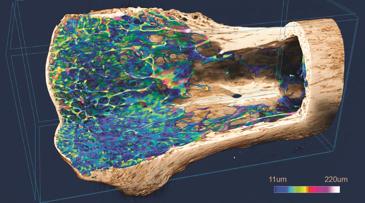
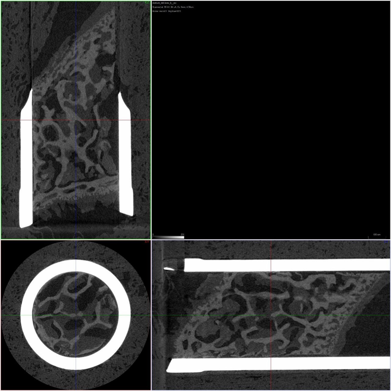
A piece of bone in a dentium drill, scanned at 4.5 µm voxel size.
3D Volume rendered image of a medieval human skull from Thuringia, scanned at 50 μm voxel size (Sample from the Friedrich-Schiller University Jena, Germany).

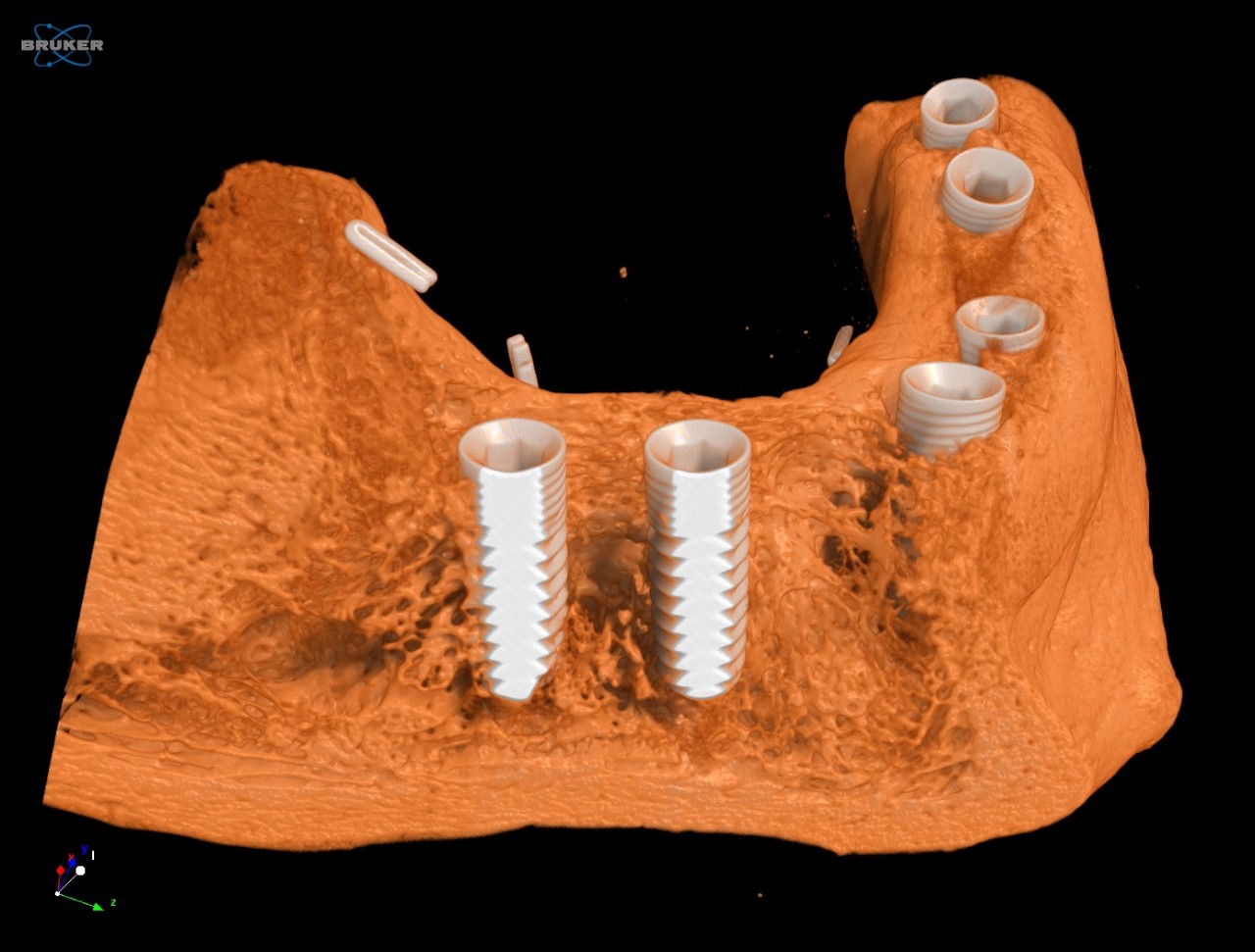
3D model of a jaw bone with several metal implants, scanned at 40 µm voxel size.
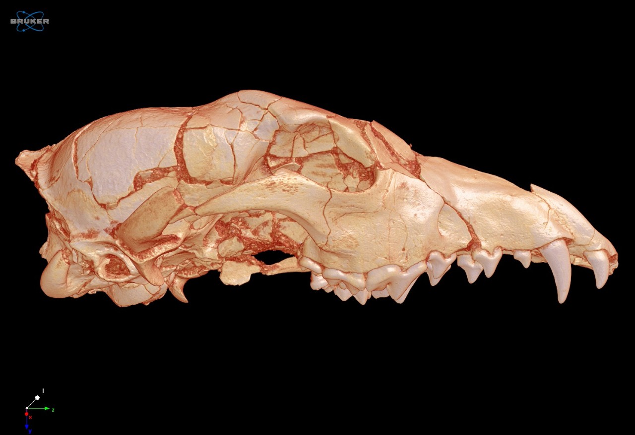
3D rendered wulf skull, scanned at 72 µm voxel size.
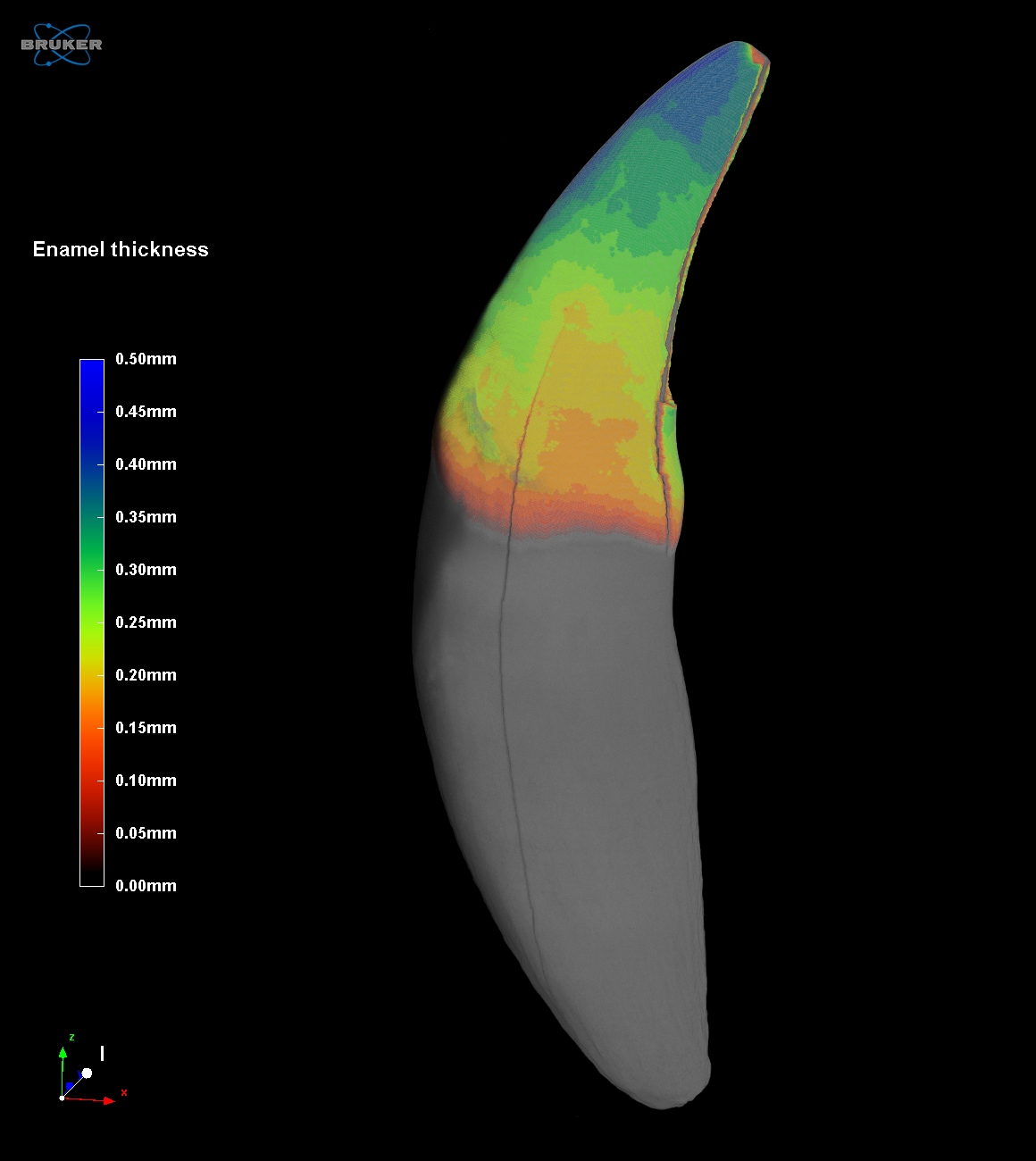
3D volume rendering of a canine tooth with color-coded representation of the enamel thickness
SKYSCAN 1273 Specifications
| Feature | Specification | Benefit |
| X-ray source | 40-130 kV | Maintenenance-free sealed X-ray source, |
| X-ray detector | Active pixel CMOS flat-panel, 6 MP (3072 x 1944) | Excellent signal-to-noise, |
| Object size | 250 mm diameter | Capability to scan huge samples in a benchtop instrument |
| Dimensions | W 1250 mm x D 815 mm x H 820 mm | Space-saving benchtop system that fits in every lab |
| Power supply | 100-240 VAC, 50-60 Hz, 3 A max. | Minimum installation requirements, a standard power supply suffices |
POSITION, SCAN, RECONSTRUCT and ANALYZE
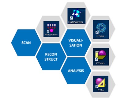
Bruker XRM solutions include all software needed to collect and analyze data. An intuitive graphical user interface with user guided parameter optimization support both expert and novice users. By using the latest GPU powered algorithms, reconstruction time is substantially reduced. CTVOX, CTAN and CTVOL combine to form a powerful suite of software for both qualitative and quantitative analysis of models.
3D.Suite Software:
Bruker microCT solutions include our comprehensive, in-house developed
3D.SUITE software for reconstruction, inspection, visualization, and
analysis of the internal object structure
Measurement Software:
SKYSCAN 2214 – Instrument control, measurement planning and collection
Reconstruction Software:
NRECON – Transforms the 2D projection images into 3D volumes
Analysis Software:
DATAVIEWER – Slice-by-slice inspection of 3D volumes and 2D/3D image registration
CTVOX – Realistic visualization by volume rendering
CTAN – 2D/3D image analysis & processing
CTVOL – Visualization of surface models to export for CAD or 3D printing
SKYSCAN 1273 - Service & Support
Available services include
- Help desk support from highly-skilled troubleshooting professionals, to isolate and resolve hard- and software problems
- Web-based remote instrument service for service diagnosis and applications support
- Merged reality support with Help Lightning – a virtual engineer at your side (video)
- Planned maintenance, according to your requirement
- Customer on-site repair and maintenance service
- Spare parts availability typically over night or within a few working days worldwide
- Compliance services for installation qualification, operational qualification / performance verification
- Site planning and relocation
Bruker Support
On our support website you will find:
Software Updates
- System software updates are available online to registered users.
Documentation
- Product manuals and installation guides are available online to registered users.
LabScape
Service & Life Cycle Support for Magnetic Resonance and Preclinical Imaging
Bruker’s commitment to provide customers with unparalleled help throughout the buying cycle, from initial inquiry to evaluation, installation, and the lifetime of the instrument is now characterized by the LabScape service concept.
LabScape Maintenance Agreements, On-Site On-Demand and Enhance Your Lab are designed to offer a new approach to maintenance and service for the modern laboratory