ParaVision 360 Highlights
Consistent Quantification
Consistent Results
ParaVision users rely on the push-button pre-optimized protocols and scan programs that come with the instrument. With CASL, Bruker debuts its first workflow package containing a complete pre-prepared scan program, an examination guide, and an integrated reconstruction with automatic measurement of inversion efficiency and output of quantitative CBF maps. This full package makes the data operator independent and therefore increases the quality and reproducibility of longitudinal studies.
Quantifiable Data
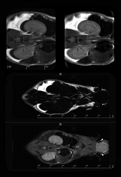
Cardiac image quality is also better than ever before. Wireless IntraGate imaging is now coupled with radial UTE to to allow 8-fold accelerated full heart cine coverage in IntraGateUTE. The minimized flow artifacts allow streamlined evaluation with automatic cardiac analysis software, and the image analysis tool allows to interactively map tissue properties.
Reproducible Results
Stability and reproducibility is a must for highly meticulous laboratories such as those of pharmaceutical companies. Scan to scan importation of parameters guarantees conformity throughout studies and subjects, and study reports ensure that all scan parameters are documented for future reference.
Efficient Animal Imaging
Animals are quickly positioned within the MRI and PET using the touchscreen of the Animal Transport System (ATS). Its accuracy guaranteers positioning correspondence between the two modalities and enables multistation acquisition for largest Field of Views.
Consistent Workflow for PET and MRI
ParaVision 360 enables full integration of PET and MRI instruments. PET/MR users profit from a commonality between workflows, image processing and analysis, and data structure and management, all of which make operation of the modalities faster and easier. Common terminology between the modalities and integrated workflows provide optimal clarity during scanning
At The Forefront of MRI
Accurate Imaging
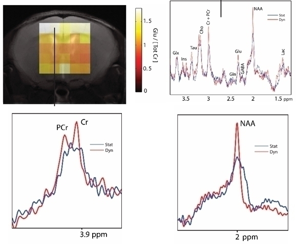
The introduction of AVANCE NEO electronics makes dynamic shimming possible, leading to even greater fidelity in fast EPI imaging and improving metabolite quantification in multi-slice CSI.
Largest Method Portfolio
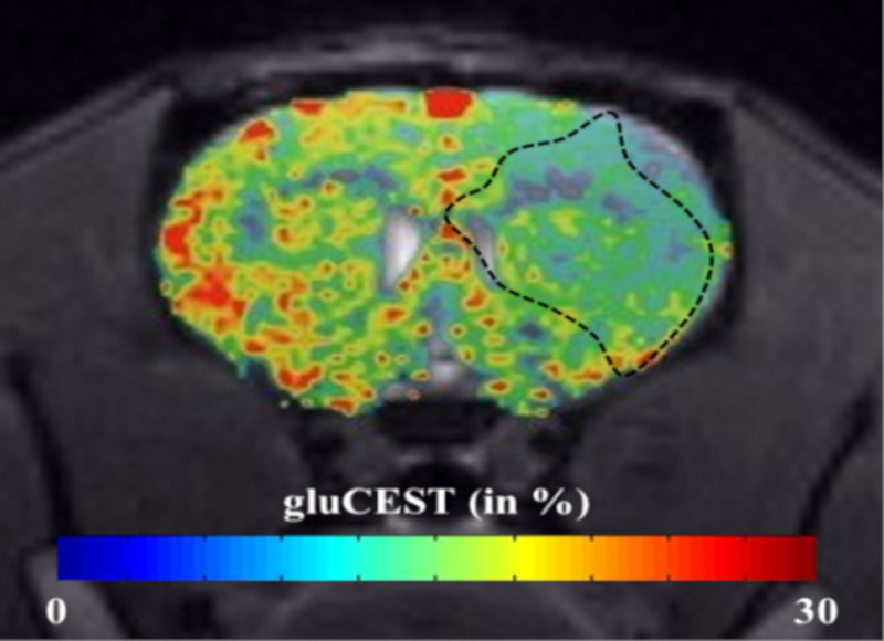
The already large MRI sequence portfolio, which is exclusive to Bruker, has been expanded even further. B1 can be mapped and shimmed based on the DREAM method and CBF can be quantitatively calculated from CASL based on EPI. The range and quality of tissue imaging is extended with the methods Double Echo Steady State (DESS), ideal for cartilage imaging and IntraGateUTE providing virtually flow artifact-free full heart cine coverage. A dual echo-time option has been added to UTE3D, and a Saturation Transfer module including CEST and MTC options is available. A fat-water separation option is provided in RARE, enabling fat chemical shift corrected images.
Powerful Data Analysis
ParaVision 360 provides extensive reconstruction, viewing and analysis functions, ranging from angles and annotation to surface rendering to underlay/overlay, to zooming/panning.
Full Flexibility
An open method developing framework with open method sources codes enables programming of novel methods and reconstruction options.
Innovative New Features
- Animal Transport System control via touchscreen
- Predefined protocols categorized via anatomy and application
- Examination guide for optimal scanning navigation, reproducibility, and consistency
- Scan program capability
- Scan to scan importation of parameters capability
- Report creation capability
- Common terminology between PET and MRI instruments
- Common integrated multi-modal workflows
- Common PET and MRI image processing, viewing, and analysis tools and functionalities
- Common PET and MRI dataset structure and management
- Automatic coregistration of PET and MRI data
- Dynamic shimming capability
- 1µs gradient resolution and synchronization
- Fast online decisions and real-time calculations on all channels
Applications
New Levels of Accuracy in Neuroscience
Once again setting the standard in preclinical imaging software, ParaVision 360 takes reproducibility to a whole new level with the introduction of the examination guide, which escorts the user throughout ASL measurements, ensuring optimal results, whether it be for investigating the extent of stroke damage and recovery or for following the progression of dementia. This consistency and reproducibility is of course only of use when the image fidelity is provided. The introduction of 1 μs gradient timing provides greatest image exactitude for best EPI quality. Additionally, dynamic shimming provides for greatest image exactitude, improving mapping of structural and functional connectivity as well as leading to more accurate quantification in CSI.
Wireless Cardiac Functional Assessment
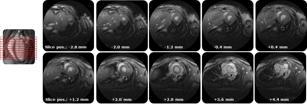
A common characteristic of cardiac models is their instability. Speed of investigation is therefore crucial for animal welfare. Valuable setup time is saved by using IntraGate, since it does not require any electrodes. This unique method that records all heart frames without the need for triggering is complimented with UTE in IntraGateUTE leading to up to 8-fold acceleration compared to classical methods. The minimized flow artifacts facilitate streamlined evaluation with automatic cardiac analysis software.
Precise Characterization of Cancer
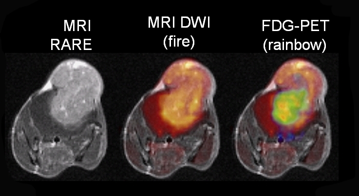
Accurate image reproducibility is imperative for oncologists measuring tumor progression and treatment. Thanks to the highest sensitivity of ParaVision 360‘s integral AVANCE NEO electronics, even smallest metastases are visible in 3D volumes. Metastases can be seen all throughout the body using multistation acquisition with subsequent image stitching. Longitudinal tumor perfusion studies benefit greatly from the ASL examination guide, which assists throughout scanning and evaluation, ensuring consistency.
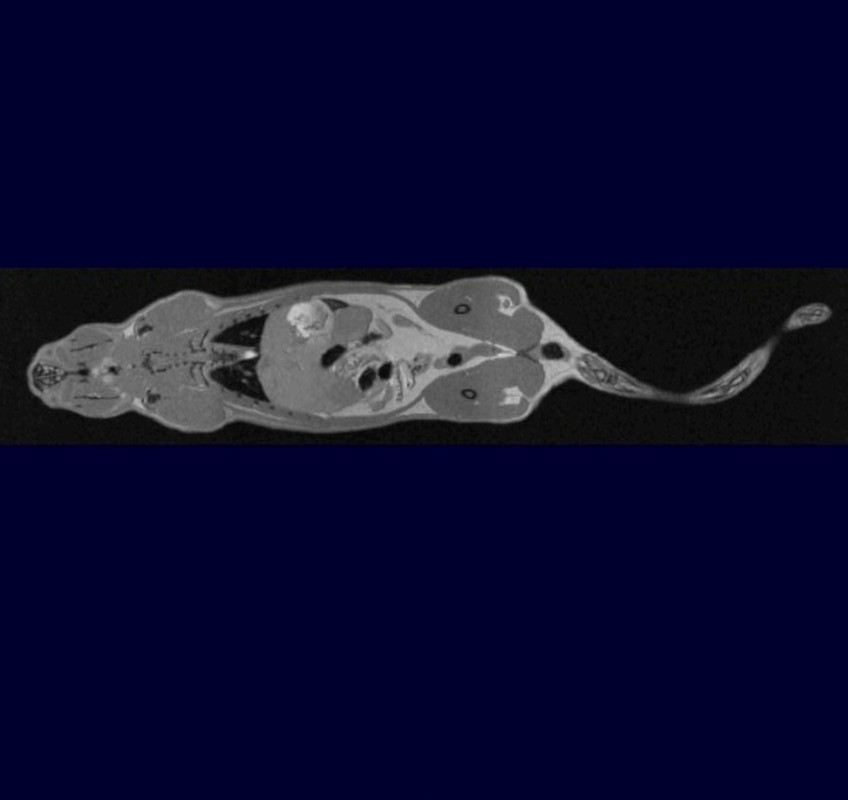
Full Body Imaging
Bruker’s largest method portfolio provides all of the methods necessary for imaging all types of tissues all throughout the body in 2D or 3D. Whether it be classic brain gray and white matter imaging with FLASH or RARE, cardiac tissue imaging with Bruker’s IntraGate FLASH or IntraGateUTE, or lung tissue or bone imaging with UTE or ZTE, Bruker’s method portfolio has you covered.
Fat and water can be imaged using the Saturation Transfer module in RARE, which includes CEST and MTC options.
Thanks to the fat chemical shift correction and to B1 shimming, image quality has never been better.
All of this imaging takes place quickly and precisely via the touchscreen positioning of the Animal Transport System (ATS). Its accuracy enables multi-station acquisition, which with subsequent image stitching yields largest Field Of Views for full body coverage. It guarantees perfect positioning correspondence between the MRI and PET images, which makes MR- based automatic attenuation correction mapping for PET possible.
Support

Service and Life Cycle Support
Bruker’s commitment to provide customers with unparalleled help throughout the buying cycle, from initial inquiry to evaluation, installation, and the lifetime of the instrument is now characterized by the LabScape service concept.
LabScape Maintenance Agreements, On-Site On-Demand and Enhance Your Lab are designed to offer a new approach to maintenance and service for the modern laboratory