Studying Texture of Paper With AFM and Upright Optical Microscopy Using the BioMAT Workstation
The BioMat Workstation enables the comprehensive structural investigation of non-transparent samples like paper. This allows researchers to gain unique insights into how nanoscale structural properties impact a material's behavior, even on samples that are otherwise inaccessible by combined AFM and optical microscopy.
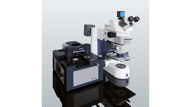
Combining AFM with optical microscopy enables detailed characterization of the surface and bulk structures of paper. Nevertheless, sample characteristics can create barriers to this method. This is particularly true for non-transparent samples, which cannot be investigated with an inverted optical microscope positioned underneath the sample. This challenge cannot be overcome through simple instrument configuration changes; in such cases, placing the objective of an upright microscope above the sample leaves too little space for the AFM to access the sample location. The BioMAT Workstation overcomes both of these problems.
Readers can expect to learn about:
- The BioMat Workstation's unique portable shuttle stage;
- How this innovative design enables the investigation of non-transparent samples;
- The full compatibility of the BioMAT Workstation with upright optical microscopy techniques; and
- Example experiments and data collected using this technical combination to investigate the structural properties of paper and toner particles.
KEYWORDS: Cellulose; Fluid Transport; Height Image; Ink Mileage; Opaque Samples; Paper;Toner Particles; Topography; Upright Optical Microscopy

Introduction
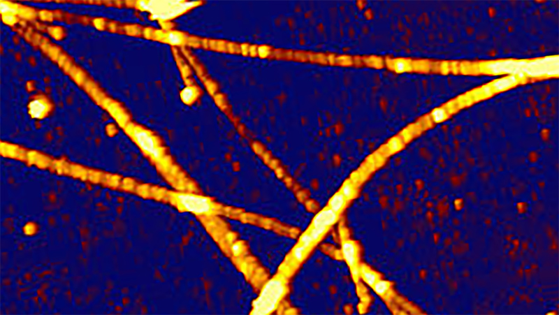
Since its development 5000 years ago in Egypt, paper is affecting our daily activities in numerous ways. It has launched a revolution in communication, packaging and personal care products, so that it is not possible to think of a world without paper – even in this digital era. In our daily lives we are familiar with different shapes and appearances of paper that are strongly connected with its capabilities. The structure of paper may influence a huge variety of characteristics like its optical, mechanical, fluid transport and print properties. In order to achieve a complete understanding of these features, a comprehensive characterization of relevant surface and bulk structures of paper is necessary (1). In Figure 1, the different appearances of two sorts of paper are presented. In general, paper is produced by pressing moist vegetable fibres of cellulose together, whereupon these macromolecules are extracted from nature products like wood, rags or grasses. Native cellulose consists of cellulose I which is a mixture of similar conformations but different lattice forms. (2) Optics and AFM together give us the ability to get a deeper insight into this multisided product and its applications.

AFM combined with upright optical microscopy
The combination of optical microscopy and AFM enable the investigation of printing and writing process on paper and how inks interact with paper. For the study of translucent samples, an inverted optical microscope can be positioned underneath the sample to combine both methods. However this does not work for paper samples which are opaque, non-transparent samples. On the other hand, placing the objective of an upright optical microscope above the sample leaves little space for the AFM to access the same location. The development of the BioMaterial (BioMAT™) Workstation overcomes these problems by using a portable shuttle stage that transports the sample between the upright microscope stage and the AFM stage
LEARN MORE:
Applications: Ink mileage
Ink mileage (or ink consumption) is a very important topic. Especially the paper and printer industries have a strong interest to create best printing results by optimal ink consumption. But also the normal end user has a concern in minimizing the concentration of potential harmful toner particles and wants to save energy and money. For perfect print results toner and surface properties of the paper are important. Especially paper porosity, pore size, permeability adhesion and roughness (3) influence the result. Lots of these properties can be measured by AFM and can be used to investigate the ink mileage. Therefore, the BioMaterial (BioMAT™) Workstation was used to combine an upright optical image with the AFM image. This ensures the ability to identify single toner particles in melted and non-melted form.


In Figure 2, the brightfield optical image and the corresponding AFM height image are shown. The combination of CMYK (Cyan, Magenta, Yellow, Black) particles allows the printing of all colors). Brightfield optical image shows intact particles and areas of molten particles. The coloured toner particle can be clearly identified in the AFM height image. Furthermore, the structural differences between the ink material and the paper surface can be distinguished from each other (cf. Figure 3).
The paper structure (Figure 3b) shows significant differences in topography and roughness in comparison to the surface of the toner particle (Figure 3c). Due to the perfect integration of optical and AFM data, the different areas can be identified in the overlay of 3D topography and brightfield optical image. Also melted and non-melted particle can be identified and assign to their colour.
Printers always aim to achieve a desired print quality with little consumption of printing ink. The ink mileage as a measure for the printing ink can be investigated easily by determining the volume of different ink particles. In Figure 4, results of such an investigation are presented, in particular the 3D topography of one toner particle and the corresponding cross-section. With the knowledge of the size and lateral expansion of the toner particle, the volume can be easily calculated. The volume of the particle that is be presented in figure 4 result in a value of 308 µm³.

Conclusion
The BioMaterial (BioMAT™) Workstation is a useful tool to combine AFM and optical imaging on opaque, nontransparent samples like paper as has been presented by the examples shown in this article. In future applications, the method could be use to investigate the ink mileage of different sorts of paper by measuring the volume of defined ink particles by AFM in combination with optical microscopy.
References
- S. J. Hanleya, J. Giassona, J.-F. Revola and D. G. Gray, Atomic force microscopy of cellulose microfibrils: comparison with transmission electron microscopy, 1992, Polymer 33 (21) , 4639-4642.
- P. Eronen, M. Österberg, U. Schmidt, and A.-S. Jääskeläinen, Effect of alkaline treatment on cellulose supramolecular
- Structure studied with combined confocal Raman
- Spectroscopy and atomic force microscopy, 2006, Colloids and Surfaces 291, 197–201.
- R. Xu A. Pekarovicova, P. D. Fleming and V. Bliznyuk Physical Properties of LWC Papers and Gravure Ink Mileage, 2005,