Revealing the PROFILE of Proteins with NMR
Monoclonal antibodies (MAbs) can be produced to recognize almost any antigen, included all cell surface and soluble receptors in the human body. As a result, their development is one of the most active areas of pharmaceutical research, as we seek to harness the considerable potential of these molecules.
Accurately characterizing the higher order structure (HOS) of protein therapeutics such as mAbs is critical, as their structure is highly related to their function. Furthermore, HOS can vary during manufacturing from lot to lot or according to the manufacturing process used, meaning that monitoring of HOS is also important for quality control purposes.
Usually, scientists characterize the 3D structure, or “fingerprint”, of a protein using 2D nuclear magnetic resonance (NMR) spectroscopy. But MAbs are such large macromolecules that the spectra produced this way have multiple overlaps and are difficult to interpret. Furthermore, glycosylation can also alter the HOS of proteins but, typically, characterizing glycosylation requires further steps such as enzymatic protein degradation and liquid chromatography-mass spectroscopy.
In a 2015 study, researchers led by Leszek Poppe showed that a 1D NMR technique called PROFILE (protein fingerprint by line shape enhancement) can simplify, speed up and increase the detail of MAb characterization.
The team first studied six lots of a glycoprotein therapeutic called epoetin alfa, which had been manufactured by two different processes (rhEPO-A and rhEPO-B). The researchers analyzed the proteins in their native, folded state to reflect the HOS, as well as in their thermally unfolded state, which reflects the sequence of amino acids in the protein.
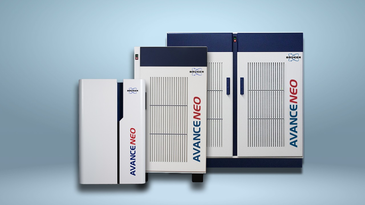
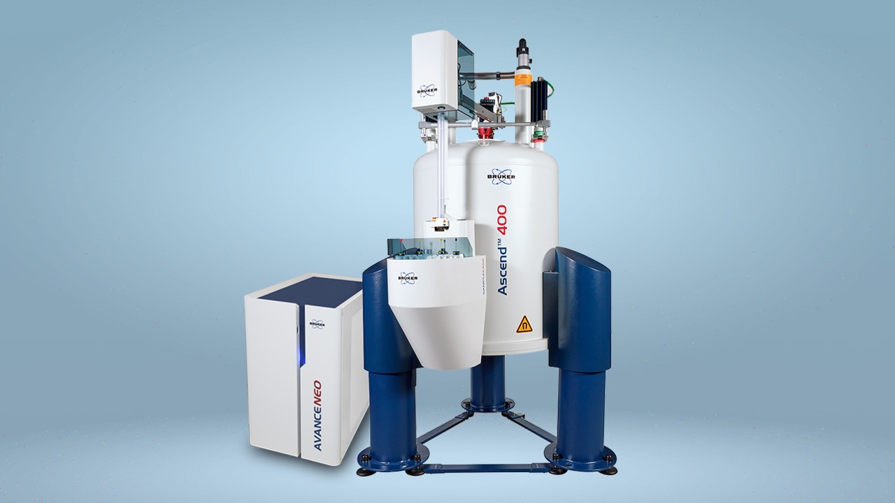
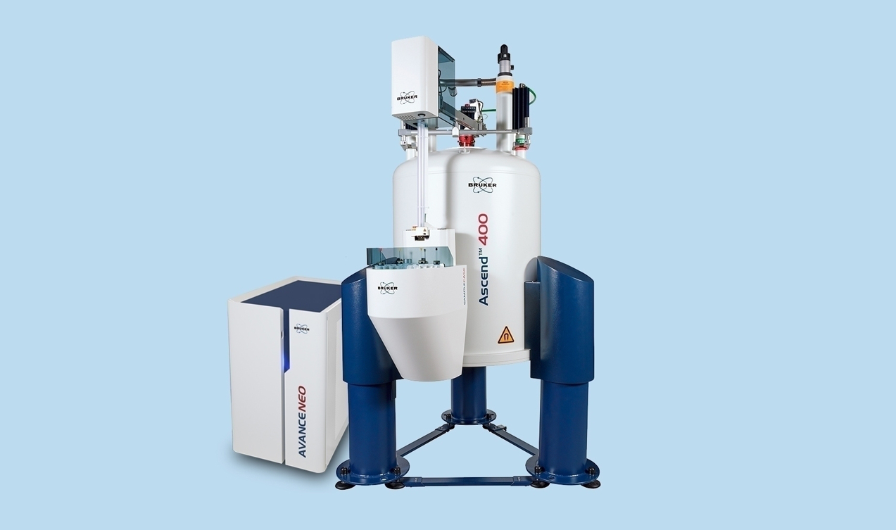
Using a Bruker Avance spectrometer, coupled with Bruker triple-resonance cryoprobes and automatic sample changer, the researchers used the PROFILE method to perform a similarity analysis. The team found that native rhEPO-A and rHEPO-B and thermally unfolded rhEPO-A and rhEPO-B were almost equally dissimilar to each other. This indicated that the differences between rhEPO-A and rhEPO-B were structural changes not attributable to HOS.
But the researchers found contrasting results when they used the method to analyze the antibody IgG1. Here they performed a similarity analysis on the glycosylated and deglycosylated forms of the antibody, in its native and unfolded states. This revealed a significant difference between the glycosylated and deglycosylated forms of the native protein, but not between the glycosylated and declycosylated forms of the unfolded protein. The only explanation for these findings is that deglycosylation of IgG1 results in significant changes to the HOS of the protein, the researchers say.
The team also showed that the PROFILE technique was much more selective than 2D NMR. Using IgG1 and IgG2 antibodies, they found that PROFILE could detect a 68% difference between the HOS of the two proteins, while 2D NMR could detect only a 42% difference. The PROFILE method was also more effective for distinguishing IgG2 molecules at low concentrations within a sample of IgG1 molecules than the 2D method.
The researchers, who report their findings in Analytical Chemistry, say that PROFILE is therefore able to provide greater information on HOS than 2D methods. They note that it also has other advantages over 2D NMR, such as that it does not require the samples to be isotopically labelled or cleaved, and can deliver faster acquisition times. The method should therefore find multiple uses in biologic pharmaceutical development and quality control, they conclude.
References
- Houde D & Engen JR. Conformational Analysis of Recombinant Monoclonal Antibodies with Hydrogen/Deuterium Exchange Mass Spectrometry. Methods Mol Biol 2013; 988: 269-289.
- Kotsovilis S & Andreakos E. Therapeutic human monoclonal antibodies in inflammatory diseases. Methods Mol Biol 2014; 1060: 37-59.
- Poppe L, Jordan JB, Lawson K, et al. Profiling Formulated Monoclonal Antibodies by 1H NMR Spectroscopy. Anal Chem 2013; 85: 9623-9629.
- Poppe L, Jordan JB, Rogers G, et al. On the Analytical Superiority of 1D NMR for Fingerprinting the Higher Order Structure of Protein Therapeutics Compared to Multidimensional NMR Methods. Anal Chem 2015; 87: 5539-5545.
- Zola H, Thomas D, Lopez A. Monoclonal Antibodies: Therapeutic Uses. eLS 2013; DOI: 10.1002/9780470015902.a0002176.pub3