Ultra High-Field Magnetic Resonance Imaging
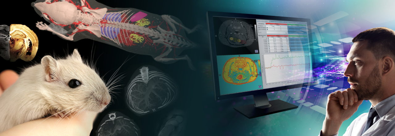
Overview
Currently, most clinical MRI systems operate at moderate field strengths of 1.5 Tesla and 3 Tesla. For small animal imaging, resolutions need to be significantly increased in order to visualize similar structures as in humans. Since the sensitivity increases with the field strength, field strengths of 7 Tesla and 9.4 Tesla are therefore standard in the preclinical field. Beyond this, preclinical UHF systems ranging from 11.7 Tesla to 18 Tesla address specific applications, which demand the highest sensitivity. Even the most demanding applications become feasible when ultra high field strengths are combined with optimal coil setups, such as receive-only arrays, the sensitivity of which increases super-linearly with magnetic field strength, or MRI CryoProbes, which provide an even additional sensitivity boost.
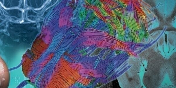
The advantages of UHF MRI go beyond the sensitivity gain itself. UHF MRI facilitates a range of imaging methods and applications. Increased chemical shift, increased Blood Oxygenation Level Dependent (BOLD) contrast, altered relaxation times, and increased susceptibility effects make it predestinated for several MRI methods such as MR Spectroscopy (MRS), BOLD Functional MRI (fMRI), Chemical Exchange Saturation Transfer (CEST), Susceptibility weighted Imaging (SWI), and Quantitative Susceptibility Mapping (QSM). Taken together UHF MRI can open up completely new avenues in the understanding of biological processes.
Increased Sensitivity
Applications at Ultra High Field (UHF) directly benefit from the high sensitivity. The gained SNR performance can, for example, be invested into higher resolutions and/or shorter scan times, or can be taken advantage of in X-nuclei imaging.
Ultra-high Resolution MRI
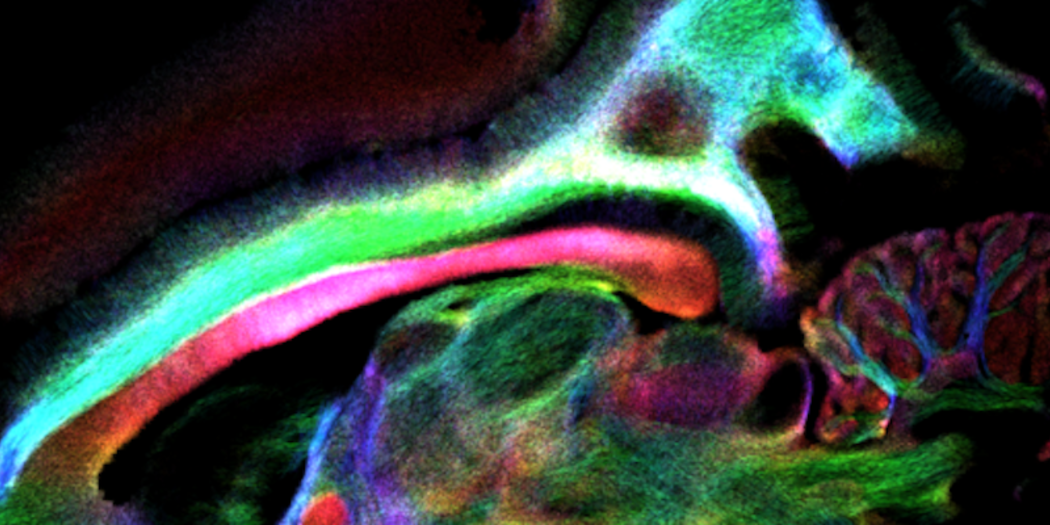
To avoid partial volume effects and thus improve data quality and data analysis, highest resolution is desired. However, if the signal in the individual voxels of the investigated subject is not large enough, the resulting low SNR prohibits analysis of the images. The greater SNR obtainable with Ultra High Field (UHF) instruments, therefore can be directly carried over into higher resolution. This enables researchers to push the resolution in the direction of “in vivo MRI histology” as well as to benefit from the increased data quality in a range of disease models [1,2].
In addition to anatomical imaging, many MRI methods benefit from the sensitivity increase. For example, in BOLD fMRI more refined stimulation paradigms can be defined, as the increased SNR places less demands on the strength of the stimulations. Furthermore, with increasing resolution, fMRI accuracy becomes less and less limited by voxel size but rather by how particularly minutely (both spatially and temporally) the blood flow to the point of neuronal activity is regulated [3]. Furthermore, for high resolution fMRI the reduced partial volume effect promises to lead to further improvement in SNR [4]. High resolution fMRI with small voxel sizes will additionally benefit from UHF since it operates in the thermal noise-dominated regime and thus, in this case, a significant sensitivity gain compared to lower magnetic fields is expected [4].

Higher Throughput

Limited available time of the MRI system, large numbers of needed animals, or instable models, often make shorter measurement times necessary.
Longer measurements times are often needed to achieve sufficient SNR when using lower field strengths. Using UHF, measurement times can be significantly shortened, since, for example, an increase in sensitivity by a factor of two allows to acquire images with the same resolution and similar quality in a quarter of the time [5]. Therefore, the number of data averages can be reduced, and the time saved can be invested into additional subjects or further studies.
A Boost for X-Nuclei Imaging
An additional benefit of the SNR gain at UHF is that imaging of X-nuclei with low gyromagnetic ratios, quadruple moments, and low abundances can be significantly improved or even made feasible for the first time [5,6,7,8].
This opens up a multitude of different research applications, such as sodium (²³Na) imaging. Sodium MRI is currently used for a wide range of applications. On clinical systems, for example, sodium concentration measurements are used to study tissue viability [9]. Due to the high sensitivity, the use of UHF greatly facilitates sodium imaging [7]. Preclinical UHF focuses, among others, on using sodium concentrations and distributions as metrics to help cellular engineers improve human mesenchymal stem cell (hMSC) conditioning for treatment of ischemic stroke [10]. Furthermore, UHF MRI can potentially lead to a breakthrough for oxygen (17O) imaging which allows direct access to the cellular oxygen metabolism. Cellular oxygen metabolism is altered in several diseases such as Alzheimer’s and Parkinson’s as well as in cancer. Thus, 17O MRI has the potential to visualize local pathology changes in the brain, underling the importance of this imaging approach [8].
Another application of X-nuclei imaging of metabolism is found with deuterium imaging, which can be used to map glucose metabolism. In addition to the ability to map glucose metabolism as opposed to glucose uptake, deuterium metabolic imaging (DMI), has the additional advantage over positron emission tomography (PET), of using non-radioactive substrates [11]. The increased sensitivity of deuterium at UHF, makes DMI a viable alternative to PET.
[1] Petiet A, Aigrot M-S, Stankoff B. Gray and White Matter Demyelination and Remyelination Detected with Multimodal Quantitative MRI Analysis at 11.7T in a Chronic Mouse Model of Multiple Sclerosis. Frontiers in Neuroscience. 2016;10:491. doi:10.3389/fnins.2016.00491.
www.ncbi.nlm.nih.gov/pmc/articles/PMC5081351/
[2] Ong HH, Webb CD, Gruen ML, Hasty AH, Gore JC, Welch EB. Fat-water MRI of a diet-induced obesity mouse model at 15.2T. Journal of Medical Imaging. 2016;3(2):026002. doi:10.1117/1.JMI.3.2.026002.
www.ncbi.nlm.nih.gov/pmc/articles/PMC4877437/
[3] Polimeni, J. and Uludağ, K., Neuroimaging with ultra-high field MRI: Present and future. NeuroImage, 2018; 168: 1-6. doi: 10.1016/j.neuroimage.2018.01.072
www.ncbi.nlm.nih.gov/pubmed/29410013
[4] Uludağ, K., and Blinder P., Linking brain vascular physiology to hemodynamic response in ultra-high field MRI. Neuroimage, 2018; 168 279-295. doi: 10.1016/j.neuroimage.2017.02.063.
www.ncbi.nlm.nih.gov/pubmed/28254456
[5] Nowogrodzki A. The world’s strongest MRI machines are pushing human imaging to new limits. Nature 563, 24-26 (2018), doi: 10.1038/d41586-018-07182-7
www.ncbi.nlm.nih.gov/pubmed/30382222
[6] Öz G, Tkáč I, Uğurbil K. Animal models and high field imaging and spectroscopy. Dialogues in Clinical Neuroscience. 2013;15(3):263-278.
www.ncbi.nlm.nih.gov/pmc/articles/PMC3811099/
[7] Deutsches Krebsforschungszentrum: www.dkfz.de/en/medphysrad/projectgroups/t7_x-nuclei/t7_x-nuclei_Na_MRI
[8] Deutsches Krebsforschungszentrum:
www.dkfz.de/en/medphysrad/projectgroups/t7_x-nuclei/t7_x-nuclei_O_MRK
[9] Thulborn KR, Lu A, Atkinson IC, Damen F, Villano J. Quantitative Sodium MR Imaging and Sodium Bioscales for the Management of Brain Tumors. Neuroimaging clinics of North America. 2009;19(4):615-624. doi:10.1016/j.nic.2009.09.001.
www.ncbi.nlm.nih.gov/pmc/articles/PMC3718497/
[10] Using High Fields to Combat Ischemic Stroke with Cell Therapy
Bruker: www.bruker.com/events/webinars/using-high-fields-to-combat-ischemic-stroke-with-cell-therapy.html
[11] De Feyter H, Behar K, Corbin Z, Fulbright R, Brown P, McIntyre S, Nixon T, Rothman D, de Graaf R. Deuterium metabolic imaging (DMI) for MRI-based 3D mapping of metabolism in vivo. Sci. Adv. 2018; 4. doi: 10.1126/sciadv.aat7314
https://advances.sciencemag.org/content/4/8/eaat7314
Higher Magnetic Susceptibility
Functional MRI (fMRI)
An application which greatly benefits from Ultra High Field (UHF) MRI is Blood Oxygenation Level Dependent (BOLD) fMRI. The increased susceptibility effects at UHF translate into a greater observable BOLD signal change and therefore improved fMRI experiments [1], as demonstrated in rat forepaw stimulation study at 15.2 Tesla, where an over 11% BOLD response was seen [2].
Functional MRI is used to study functional connectivity to further understand brain function in health and disease [3]. Using the high sensitivity provided by UHF, high resolution fMRI preclinical experiments thus become feasible [4]. Forepaw somatosensory stimulation, for example, only commonly shows BOLD response in S1FL. A recent study at 9.4 T and 15.2 T, however, detected only S1FL response at 9.4 T, but S2 and thalamus as well as S1FL at 15.2 T [5]. Functional sensitivity will additionally benefit from UHF in situations where thermal noise is dominant, as it is directly dependent on sensitivity and indirectly dependent on temporal noise [2]. This is the case for high resolution studies which are enabled at UHF [6].

SWI and QSM

In addition to BOLD imaging, further imaging applications which rely on high susceptibility effects combined with a high SNR and therefore benefit from UHF, are Susceptibility Weighted Imaging (SWI) and Quantitative Susceptibility Mapping (QSM) [7]. QSM can for example be applied in vivo to study the microvasculature in animal stroke models [8].
[1] Duyn JH. The future of ultra-high field MRI and fMRI for study of the human brain. Neuroimage. 2012;62(2):1241-1248. doi:10.1016/j.neuroimage.2011.10.065.
www.ncbi.nlm.nih.gov/pmc/articles/PMC3389184/
[2] Han SH, Son JP, Cho HJ, Park JY, Kim SG. Gradient‐echo and spin‐echo blood oxygenation level–dependent functional MRI at ultrahigh fields of 9.4 and 15.2 Tesla. Magnetic Resonance in Medicine. 2018; 1-10. doi: 10.1002/mrm.27457 www.ncbi.nlm.nih.gov/pubmed/30183108
[3] Kalthoff D, Hoehn H. Functional Connectivity MRI of the Rat Brain. Bruker Application Note 2012
Functional_Connectivity.pdf
[4] Seehafer JU, Hoehn H. Insights in the rat brain by high resolution BOLD functional MRI. Bruker Application Note 2011 BOLD_MRI_AppsNote_T13106.pdf
[5] Jung WB, Shim HJ, Kim SG. Mouse BOLD fMRI at ultrahigh field detects somatosensory networks including thalamic nuclei. NeuroImage. 2019;195: 203-214. doi.org/10.1016/j.neuroimage.2019.03.063 www.ncbi.nlm.nih.gov/pubmed/30946950
[6] Uludağ K, Blinder P. Linking brain vascular physiology to hemodynamic response in ultra-high field MRI. NeuroImage. 2018; 168: 279-295. doi.org/10.1016/j.neuroimage.2017.02.063 www.ncbi.nlm.nih.gov/pubmed/28254456
[7] Duyn J. MR Susceptibility Imaging. Journal of magnetic resonance (San Diego, Calif : 1997). 2013;229:198-207. doi:10.1016/j.jmr.2012.11.013.
www.ncbi.nlm.nih.gov/pmc/articles/PMC3602381/
[8] Hsieh M-C, Tsai C-Y, Liao M-C, Yang J-L, Su C-H, Chen J-H. Quantitative Susceptibility Mapping-Based Microscopy of Magnetic Resonance Venography (QSM-mMRV) for In Vivo Morphologically and Functionally Assessing Cerebromicrovasculature in Rat Stroke Model. Jiang Q, ed. PLoS ONE. 2016;11(3):e0149602. doi:10.1371/journal.pone.0149602.
www.ncbi.nlm.nih.gov/pubmed/26974842
Higher Spectral Dispersion
Spectroscopy
Due to the increased sensitivity and high spectral dispersion at Ultra High Field (UHF), a natural UHF application is magnetic resonance spectroscopy (MRS). Commercially available MRS instruments are readily available today with field strengths up to 28 Tesla, allowing ultra-high resolution spectroscopy experiments of small samples [1].
Similarly, UHF MRI magnets can exploit the increased chemical shift and sensitivity in vivo. Thus, significant improvements in preclinical in vivo MRS have been reported when using UHF magnets [Öz 2013, Shemesh Nat Commun 2014, Mlynárik 2008, Shemesh Journal of Cerebral Blood Flow 2014]. Whereas spectrally edited sequences, such as MEGA-PRESS, allow GABA imaging at lower field strengths, straight-forward sequences can be used at UHF. For example, at 15.2 T PRESS was used for fMRS on GABA in chemogenetically engineered mice. [Zohar 2019] Notably, it has been shown that certain metabolites could be detected for the first time in vivo due to the high magnetic field strength [Mlynárik 2008].
Beyond the benefits in sensitivity gain and high chemical shift, it could be further shown that a relaxation enhanced MRS strategy allows to additionally utilize the difference in relaxation times between water and metabolites at UHF to generate “water-free” MRS without the need for common water suppression techniques [Shemesh Nat Commun 2014].

Chemical Exchange MRI
In addition to spectroscopy, the increased spectral dispersion also benefits magnetization transfer techniques such as Chemical Exchange Saturation Transfer (CEST) imaging, leading to a high selectivity [Bruker CEST]. Further advantages of chemical exchange techniques at UHF include the higher saturation which can be achieved [Bruker CEST] and a reduction of the exchange rate relative to the chemical shift [Chung 2017]. As the exchange rate must be smaller than the chemical shift, the increased spectral dispersion allows for faster exchanging compounds to be detected [Wu 2016].
To ascertain optimal conditions at preclinical as well as clincal field strengths, McMahon et al. conducted simulations of chemical shift, exchange rate, and detection sensitivity and determined a significantly larger usable chemical shift range at 11.7 T than at 3 T. [BrukerCEST 2021]
A recent paper from Chung et al. demonstrated a significantly increased chemical exchange effect for the amine proton signal in rat brain at 15.2 Tesla compared to 9.4 Tesla. An increase of 65% compared to 9.4 Tesla was reported emphasizing the importance of UHF for chemical exchange applications [Wu 2016, Bruker fMRI]. The same group also investigated phosphocreatine (PCr) modulation with PCrCEST in the hindlimb of mice and found a 29% higher PCrCEST signal at 15.2 Tesla than at 9.4 Tesla. The significant sensitivity of PCrCEST in hindlimb indicates that PCrCEST could be valuable for mapping energy metabolism in muscles such as the heart [Chung 2019].
A prominent CEST application which benefits from UHF is GluCEST, which monitors local metabolic defects in neurodegenerative diseases [Bruker CEST, Pépin 2016]. For example, GluCEST was identified in the past to be a potential in vivo biomarker of Huntington’s disease when applied in a knock-in mouse model using UHF [Pépin 2016], and has recently been used in a mouse epilepsy model, in which chronically methionine sulfoximine (MSO)-seizure-inducing treated mice showed reduced GluCEST contrast in the hippocampus [Bagga 2019].
Furthermore, there are indications that glucoCEST can be used to investigate metabolism associated with neuronal activity. GlucoCEST performed at 17.2 Tesla using a rat model of electrical forepaw stimulation showed negative contrast during stimulation in the same regions that BOLD imaging provided positive contrast, thus demonstrating the ability of CEST fMRI to locally monitor temporal changes of glucose concentration [Roussel 2019].

[1] Overview Ascend GHZ Class
[2] Öz G, Tkáč I, Uğurbil K. Animal models and high field imaging and spectroscopy. Dialogues in Clinical Neuroscience. 2013;15(3):263-278.
www.ncbi.nlm.nih.gov/pmc/articles/PMC3811099/
[3] Shemesh N, Rosenberg JT, Dumez J-N, Muniz JA, Grant SC, Frydman L. Metabolic properties in stroked rats revealed by relaxation-enhanced magnetic resonance spectroscopy at ultrahigh fields. Nat Commun. 2014; 5: 4958. doi: 10.1038/ncomms5958
www.ncbi.nlm.nih.gov/pubmed/25229942
[4] Mlynárik V, Cudalbu C, Xin L, Gruetter R. 1H NMR spectroscopy of rat brain in vivo at 14.1Tesla: improvements in quantification of the neurochemical profile. J Magn Reson. 2008; 194: 163–168. doi: 10.1016/j.jmr.2008.06.019
www.ncbi.nlm.nih.gov/pubmed/18703364
[5] Shemesh N, Rosenberg JT, Dumez J-N, Grant SC, Frydman L. Metabolic T1 dynamics and longitudinal relaxation enhancement in vivo at ultrahigh magnetic fields on ischemia. Journal of Cerebral Blood Flow 2014;34(11):1810-1817. doi:10.1038/jcbfm.2014.149.
www.ncbi.nlm.nih.gov/pmc/articles/PMC4269758/
[6] Zohar I, Saraf-Sinik I, Yizhar O, Tal A. Functional magnetic resonance spectroscopy towards detection of initiated release of GABA in chemogenetically engineered mice. ISMRM 2019 2233
https://index.mirasmart.com/ISMRM2019/PDFfiles/2233.html
[7] Metabolic Imaging in Neurodegenerative Disease using CEST MRI | Bruker: www.bruker.com/service/education-training/webinars/pci-webinars
[8] Chung JJ, Choi W, Jin T, Lee JH, Kim S-G. Chemical-exchange-sensitive MRI of amide, amine and NOE at 9.4 T versus 15.2 T. NMR in Biomedicine. 2017;30:e3740. doi.org/10.1002/nbm.3740
www.ncbi.nlm.nih.gov/pubmed/28544035
[9] Wu B, Warnock G, Zaiss M, Lin C, Chen M, Zhou Z, Mu L, Nanz D, Tuura R, Delso G. An overview of CEST MRI for non-MR physicists. 2016; EJNMMI Physics 3:19
www.ncbi.nlm.nih.gov/pubmed/27562024
[10] New insights into brain function with molecular and functional MRI of the rodent brain at ultra-high fields | Bruker: www.bruker.com/service/education-training/webinars/pci-webinars
[11] Chung JJ, Jin T, Lee JH, Kim SG. Chemical exchange saturation transfer imaging of phosphocreatine in the muscle. Magnetic Resonance in Medicine 2019, 81(6):3476-3487. doi: 10.1002/mrm.27655
www.ncbi.nlm.nih.gov/pubmed/30687942
[12] Pépin J, Francelle L, Carrillo-de Sauvage M-A, de Longprez L, Gipchtein P, Cambon K, Valette J, Brouillet E, Flament J. In vivo imaging of brain glutamate defects in a knock-in mouse model of Huntington's disease. Neuroimage. 2016; 139: 53–64. doi: 10.1016/j.neuroimage.2016.06.023
www.ncbi.nlm.nih.gov/pubmed/27318215
[13] Bagga P, Pickup S, Flament J, Detre J, Hariharan H, Reddy R. Mapping astroglial glutamine synthetase activity in vivo in a preclinical model of epilepsy using glutamate-weighted CEST (GluCEST) MRI. ISMRM 2019 3121
https://index.mirasmart.com/ISMRM2019/PDFfiles/3121.html
[14] Roussel T, Frydman L, Le Bihan D, Ciobanu L. Brain sugar consumption during neuronal activation detected by CEST functional MRI at ultra-high magnetic fields. Scientific Reports 9(1):4423. doi.org/10.1038/s41598-019-40986-9 1
www.ncbi.nlm.nih.gov/pubmed/30872689
[Bruker CEST 2021] Characteristics of Organic Compounds Producing Strong CEST Contrast and How to Use These For Cancer and Kidney Imaging | Bruker: www.bruker.com/service/education-training/webinars/pci-webinars
Phased Arrays and MRI CryoProbes
Receive Arrays
For clinical systems, it was recently shown that for receive-only arrays the Signal-to-Noise-Ratios (SNR) increases super-linearly with the magnetic field strength [1]. These results underline the impressive SNR gain when moving to Ultra High Field (UHF) systems.
Besides the SNR gain itself, receive arrays coils further provide acceleration possibilities, thus having the potential to increase spatial as well as temporal resolution. For preclinical UHF imaging, array coils are available for brain, heart, spine and body and have found routine use.
MRI CryoProbes

In preclinical MRI, cryogenically-cooled MRI CryoProbes [2], provide an additional sensitivity boost [3] and have found widespread use. Combined with UHF MRI, the additional gain is significant and enables highest quality images in reasonable measurement times [4]. Thus, for example, ultra high resolution in vivo mouse brain data can be easily acquired on a preclinical 15.2 Tesla equipped with a MRI CryoProbe.
[1] Pohmann, R, Speck, O, Scheffler, K. Signal-to-noise ratio and MR tissue parameters in human brain imaging at 3, 7, and 9.4 tesla using current receive coil arrays. Magn. Reson. Med., 2016; 75: 801-809. doi:10.1002/mrm.25677
www.ncbi.nlm.nih.gov/pubmed/25820458
[2] MRI CryoProbes | Bruker www.bruker.com/products/mr/preclinical-mri/mri-cryoprobes.
[3] Niendorf T, Pohlmann A, Reimann HM, et al. Advancing Cardiovascular, Neurovascular, and Renal Magnetic Resonance Imaging in Small Rodents Using Cryogenic Radiofrequency Coil Technology. Frontiers in Pharmacology. 2015;6:255. doi:10.3389/fphar.2015.00255.
www.ncbi.nlm.nih.gov/pmc/articles/PMC4642111/
[4] Petiet A, Aigrot M-S, Stankoff B. Gray and White Matter Demyelination and Remyelination Detected with Multimodal Quantitative MRI Analysis at 11.7T in a Chronic Mouse Model of Multiple Sclerosis. Frontiers in Neuroscience. 2016;10:491. doi:10.3389/fnins.2016.00491.
www.ncbi.nlm.nih.gov/pmc/articles/PMC5081351/


Additional information
LabScape
Service & Life Cycle Support for Magnetic Resonance and Preclinical Imaging
Bruker’s commitment to provide customers with unparalleled help throughout the buying cycle, from initial inquiry to evaluation, installation, and the lifetime of the instrument is now characterized by the LabScape service concept.
LabScape Maintenance Agreements, On-Site On-Demand and Enhance Your Lab are designed to offer a new approach to maintenance and service for the modern laboratory