What is Cleared-Sample Imaging?
Clearing-Sample/ Cleared-Tissue Imaging
Tissue clearing techniques have become a valuable tool for applications in 3D microstructure analysis of tissues (e.g. neuroscience, developmental biology, connectomics).
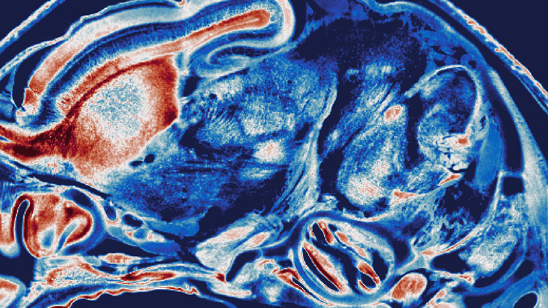
The different refractive indexes (RI) of the major components of biological tissue, i.e. water, lipids and proteins result in light scattering when light passes through the tissue. Tissue clearing modifies the optical properties of usually opaque samples to render them transparent while keeping their structure and fluorescent labels intact. After clearing, light can travel many millimeters through a specimen unrestricted from absorption and scattering, ideal for high-resolution microscopic imaging deep within the specimen.
Light-Sheet Microscopy leverages the optical advantages of cleared samples enabling fast, long-term, confocal-like optical sectioning and high-quality 3D imaging of cleared samples.

Methods for Optical Clearing
There is a huge variety of different clearing methods, and answering which of the available clearing techniques is best for your specific sample is not easy as it mainly depends on the type of tissue, the labeling available/used, the sample size, etc.
However, the clearing techniques can be categorized into 3 major classes:
A recent categorization chose the naming: Hydrophobic, hydrophilic, hydrogel-based.
Another common naming scheme is: Solvent based, aqueous based (including hyperhydration), and hydrogel embedding.
Examples of hydrophobic clearing methods: BABB, DISCO-family, Visikol, ECi, PEGASOS, DBE, etc.
Examples of hydrophilic clearing methods: Scale-family, CUBIC-family, FRUIT, SeeDB, FocusClear, ClearT, ClearSee, Ce3D, OPTIclear, DEEP-clear, MACS, etc.
Examples of hydrogel-based clearing methods: CLARITY, PACT, PARS, SWITCH, SHIELD, etc.
FLASH, a customizable technique for molecular phenotyping, can be used with a variety of clearing protocols from different classes, such that it can be adapted to a wide range of different tissues.