Super-Resolution Microscopy Protocols
Provided by Bruker super-resolution microscopy experts.
Sample Preparation Protocols
Fluorescent Labeling Protocols
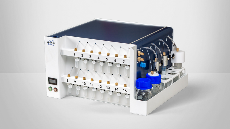
Buffer and Material Preparation Protocols
Staining Protocols
The standard procedures and protocols for SMLM methods are provided here.
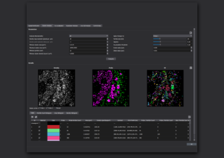
Single-molecule localization microscopy (SMLM) enables the observation, quantification, and tracking of many cellular structures and molecular structures. Moreover, SMLM techniques (e.g., dSTORM, PALM, DNA-PAINT, and OligoSTORM) provide previously unobtainable data, like the composition of synapses and the interaction of viruses with sub-cellular structures.
The optical system, analysis software, dye/fluorophore selected, and other experimental parameters will impact the achievable spatial and temporal resolution. Such selections also determine whether the technique can be used to image fixed or live cells, acquire images in two or three dimensions, or perform single- or multi-color localization. Key adjustable parameters include:
- Sample preparation (e.g., fixation method and buffer used);
- Labeling strategies; and
- Staining methods.