
肺病学
肺部的临床前成像推动了呼吸系统疾病的研究
肺部成像推动我们对呼吸系统疾病的了解
肺病学是一门预防、诊断和治疗那些影响呼吸系统疾病的科学。除了如哮喘和慢性阻塞性肺疾病这样的呼吸系统疾病,还有一系列其他慢性疾病会对肺部造成影响,如心血管疾病或癌症。肺部成像能使专业医疗卫生人员进一步了解肺部从其他一系列疾病上所受到的影响,从而优化治疗方案。
成像手段可帮助研究人员确定许多肺部疾病的确切病因,这可显著提升肺病学诊断的准确性。临床前小动物成像尤为重要——可为肺部疾病的机理以及治疗效果提供深入分析。
此类基于计算机断层扫描、磁共振成像以及正电子发射断层扫描的研究已测定出一些关键数据。这些数据可确定肺部疾病的诊断,如肺炎、肺栓塞或肺动脉高压、肺部肿瘤的早期检测和阶段性支气管癌等。
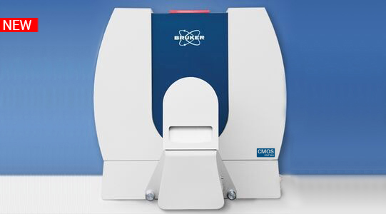
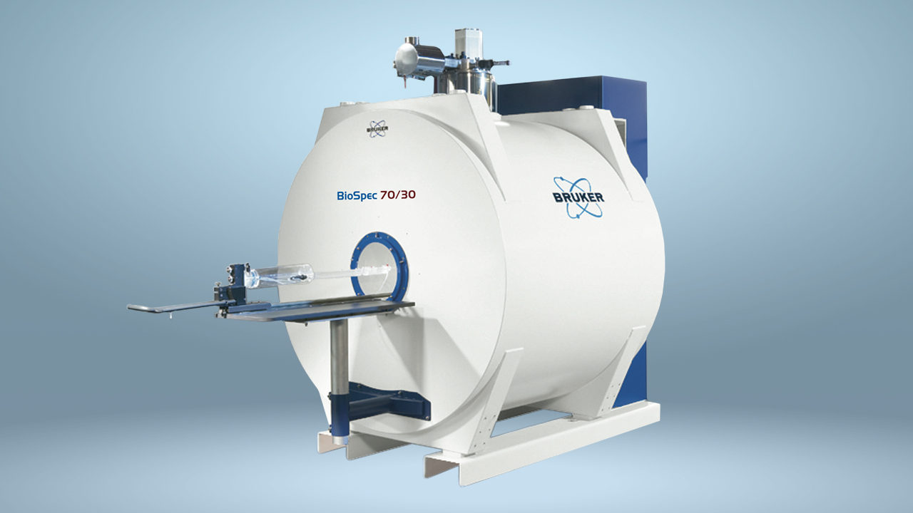
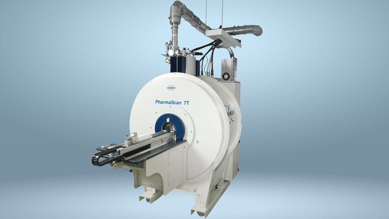
布鲁克丰富的分子成像技术组合,包括肺部PET与MR整合的系统,以及可用于活体动物研究的micro-CT扫描仪。

支持

服务和生命周期支持
布鲁克致力于在整个购买周期内为用户提供出色的帮助,从最初的咨询到评估、安装以及仪器的全使用周期,现在均包含在LabScape服务理念当中。
LabScape维保协议、现场按需服务和实验室升级服务,旨在为现代实验室提供一种全新的维护和服务方式。