Advancing the Development of Drugs
Preclinical Imaging – Gain crucial knowledge at an early stage
Preclinical imaging plays a crucial role in understanding how the body works in both healthy and disease states and describing responses to physiological or environmental change. It provides important insights into disease mechanisms at organ, tissue, cell, and molecular levels.
Such knowledge informs the development of novel therapeutic strategies, which in turn improve patient outcomes and save lives. Preclinical imaging is also central to evaluating the effectiveness and safety of new treatments and describing drug distribution patterns before clinical use.
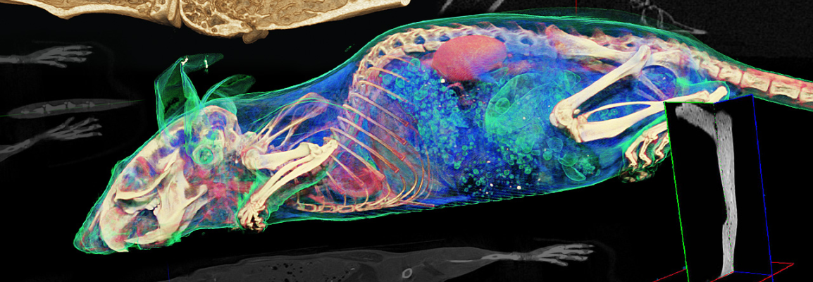
Cost-efficient and high-throughput longitudinal study of animal models can be achieved using anatomical evaluation techniques such as magnetic resonance imaging (MRI) and computed tomography (CT) alongside positron emission tomography (PET) and single-photon emission computed tomography (SPECT) for molecular visualizations.
Bruker has developed a wide portfolio of molecular imaging technologies to meet varying preclinical imaging requirements. It includes MRI and CT scanners specifically designed for in vivo rodent imaging, such as the 9.4 Tesla (T) small animal MRI scanner and the MicroCT scanner. Multimodal imaging is facilitated by the combination of technologies in a single, easy-to-use instrument, such as the Albira system that includes PET, SPECT and CT technologies and the PET/MR 3T that combines PET and MRI.