Imaging Techniques for Assessing the Effects of Cerebral Ischaemia In Vivo
Cerebral ischemia
Cerebral ischemia is the most common cause of stroke, which is a leading cause of permanent disability in adults worldwide1,2 It occurs when blood vessels serving the brain become blocked or burst depriving it of sufficient oxygen and nutrients. Consequently, the affected area of the brain cannot work properly and the bodily functions it controls are impaired, giving rise to a variety of disabilities, such as reduced mobility and coordination and impaired speech.
Despite advances in the understanding of the pathophysiology of cerebral ischemia, there are few therapeutic options2. The majority of treatments are centered on reperfusion of the affected area of the brain to restore the supply of oxygen to the deprived cells and enable normal function to resume as quickly as possible.
Although several different mechanisms are involved in the pathogenesis of cerebral ischemia, the resultant inflammation of brain tissue is believed to be a key factor in determining the extent of irreversible brain damage and associated disability3. Increases in the levels of inflammatory markers have been found to correlate with poor outcome after cerebral ischemia. The molecular basis of the inflammatory response to cerebral ischemia is poorly understood, but a role of nicotinic acetylcholine receptors (nAChRs) has been indicated4.
Nicotinic acetylcholine receptors
The ligand-gated nAChRs are ion channels that are widely expressed throughout the peripheral and central nervous systems. A subtype of this group of receptors known as α7 is present across a range of different brain cells, including cortical neurons, microglia and astrocytes, where it serves a vital role in the control of a wide variety of physiological responses such as attention, memory, and locomotion5.
The α7 receptors have been implicated in the development of schizophrenia, Alzheimer’s disease and traumatic brain injury. Furthermore, stimulation of α7 receptors appears to provide neuronal protection against ischemic damage and cerebral haemorrhage6,7.
Further elucidation of the function of these receptors may thus help reduce cerebral inflammation and improve outcomes in patients with cerebral ischemia and brain diseases. To investigate the role of these receptors, it is necessary to study them in vivo, and sophisticated imaging techniques have now made this possible.
Cerebral imaging
Positron emission tomography (PET) imaging and magnetic resonance imaging (MRI) are powerful tools for visualizing the effects of neurological and neurodegenerative diseases in vivo. Since these imaging techniques do not interfere with the normal functioning of the body they can be repeated several times to assess effects over time.
Selective PET radiotracers specific for α7 receptors have allowed the distribution of these receptors to be visualized. Although such studies have highlighted the role of α7 nAChRs in brain disorders, the role of nAChRs in the neuroinflammatory response to cerebral ischemia had not been determined.
Role of nicotinic receptors in cerebral ischemia
To rectify this gap in the understanding of nAChRs involvement in cerebral ischemia, changes in the expression of the α7 receptor were studied in a rat model after inducing cerebral ischemia in the brain by a 90-minute intraluminal occlusion of the middle cerebral artery8.
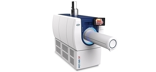
Rats with cerebral ischemia were treated with the α7 nAChRs agonist PHA 568487 or control and the effects visualized by in vivo PET imaging with [11C]NS14492 as the radiotracer. MRI was conducted using a 7 Tesla horizontal bore Bruker BioSpec 70/30 MRI system to monitor permeability of the blood brain barrier. The effect of the modulation of α7 receptors on neuroinflammation was explored with a specific radioligand for the translocator protein ([18F]DPA-714) 7 days after cerebral ischaemia. Real-time polymerase chain reaction was used to evaluate changes in gene expression.
Increased expression of α7 receptors was observed in microglia and astrocytes after cerebral ischaemia. The role of α7 receptors in the neuroinflammatory response to cerebral ischemia was supported by the decrease of [18F]DPA-714 binding in ischemic rats treated with PHA 568487. In addition, treatment with PHA 568487 significantly reduced cerebral infarct volumes and improved neurological outcomes compared with control. Activation of α7 nAChRs had no influence on blood brain barrier permeability8.
Taken together, these results suggest that the nicotinic α7 nAChRs play a key role in the inflammatory reaction and the leukocyte recruitment following cerebral ischemia in rats
It is hoped that the new information obtained in this study supporting the involvement of α7 nAChRs in the development of neuroinflammation after cerebral ischemia may facilitate the development of novel strategies to reduce disability after ischemic stroke and treatments to reduce the burden of other neurological diseases.
References
1. Anuncibay-Soto B, et al. Neuroprotection by salubrinal treatment in global cerebral ischemia. Neural Regen Res 2016;11:1744‑1745.
2. Donnan GA, et al. Stroke. Lancet. 2008, 371: 1612‑1623. .
3. Muir KW, et al. Inflammation and ischaemic stroke. Curr Opin Neurol. 2007;20:334‑342.
4. de Jonge WJ and Ulloa L. The alpha7 nicotinic acetylcholine receptor as a pharmacological target for inflammation. British Journal of Pharmacology 2007;151(7), 915–929.
5. Neumann S, et al. Innate immunity and inflammation post-stroke: An alpha7-nicotinic agonist perspective. International Journal of Molecular Sciences 2015;16(12),29029–29046.
6. Shimohama S, et al. Nicotinic alpha 7 receptors protect against glutamate neurotoxicity and neuronal ischemic damage. Brain Research1998;779:359–363.
7. Hijioka M, et al. Therapeutic effect of nicotine in a mouse model of intracerebral hemorrhage. Journal of Pharmacology and Experimental Therapeutics 2011;338(3):741–749.
8. Colás L, et al. In vivo imaging of A7 nicotinic receptors as a novel method to monitor neuroinflammation after cerebral ischemia. Glia 2018;1–14. Available here.

Support

Service and Life Cycle Support
Bruker’s commitment to provide customers with unparalleled help throughout the buying cycle, from initial inquiry to evaluation, installation, and the lifetime of the instrument is now characterized by the LabScape service concept.
LabScape Maintenance Agreements, On-Site On-Demand and Enhance Your Lab are designed to offer a new approach to maintenance and service for the modern laboratory