Episode 1: Interview with an AFM Pioneer: Prof. Mervyn Miles
Episode 1: Interview with an AFM Pioneer
In this episode of the Conversations on AFM podcast series, we speak to Prof Mervyn Miles, a pioneer in the field of AFM. Prof Miles talks about the technology, placing it in a historical context, from the early beginnings to Dr. Toshio Ando’s groundbreaking work on the development of high-speed AFM. He speaks on trends and applications, from the importance of automation, high-speed data acquisition, and AI machine learning to future applications in biomedical science.
TO LEARN MORE:
Conversations on AFM: Exploring Its Origins and Impact Today
Atomic Force Microscopy (AFM) is a microscopy technique that has revolutionised our understanding of materials at the nanoscale. Developed in the mid-1980s, AFM uses a probe to scan over a sample to describe its surface topography and other properties, such as tissue stiffness or temperature. Unlike conventional imaging techniques, which often rely on light or magnetic fields, AFM measures probe-to-surface interactions, offering unparalleled detail at the atomic level.
AFM is as inspiring as it is complex, so we created the “Conversations on AFM” Bruker podcast. In the first episode, we interviewed the AFM pioneer Emeritus Professor Mervyn Miles from the University of Bristol, also a co-founder of two spin-out companies in AFM, to learn more about the history of AFM.
How Does AFM Work?
AFM functions by scanning a sample with a small tip, often just a few nanometers in diameter, across the surface of a sample. The probe is mounted on a flexible cantilever, which deflects in response to interactions between the probe and the surface. As the probe moves over the sample, various forces — such as van der Waals, electrostatic, and mechanical forces — affect the cantilever's deflection. By measuring these deflections with high precision, AFM can map the topography and mechanical properties of the surface [1].
What Makes AFM Special?
AFM's ability to operate in different environments, including air, vacuum, and liquid, makes it an invaluable tool for a wide range of applications. This adaptability allows researchers to study polymers, metals, semiconductors, and biological samples under conditions that closely mimic their natural states.
The Legacy of AFM Pioneers
Scanning Probe Microscopy (SPM) is a collective term for AFM and Scanning Tunnelling Microscopy. For the latter, the 1986 Nobel Prize in Physics was awarded to Gerd Binnig and Heinrich Rohrer. Prof Dr Gerd Binnig went on to develop AFM with Calvin Quate and Christoph Gerber, extending the concept of STM to non-conductive surfaces. This allowed AFM to be applied to a wider range of materials beyond conductors, making AFM invaluable in many scientific fields, especially in biology, chemistry, and materials science.
In our AFM podcast with Prof Miles, he shared that the development of AFM was not only groundbreaking because it relied on mechanics and eliminated the need for samples to be conductive but also because it overcame the resolution limitations of optical microscopes. This combination had profound implications for applying AFM in life science research, enabling researchers to study biomolecular structures and processes without altering the natural state of the sample.
Applications of BioAFM
Prof Miles also shared that BioAFM, a variant of AFM tailored for biological samples, provides insights into cellular rigidity and the mechanical properties of biological tissues, which can be crucial for medical diagnostics. This ability to investigate living cells and tissues in their natural environments opens new avenues for understanding disease mechanisms and developing therapeutic strategies [2].
BioAFM Application 1: Measuring DNA
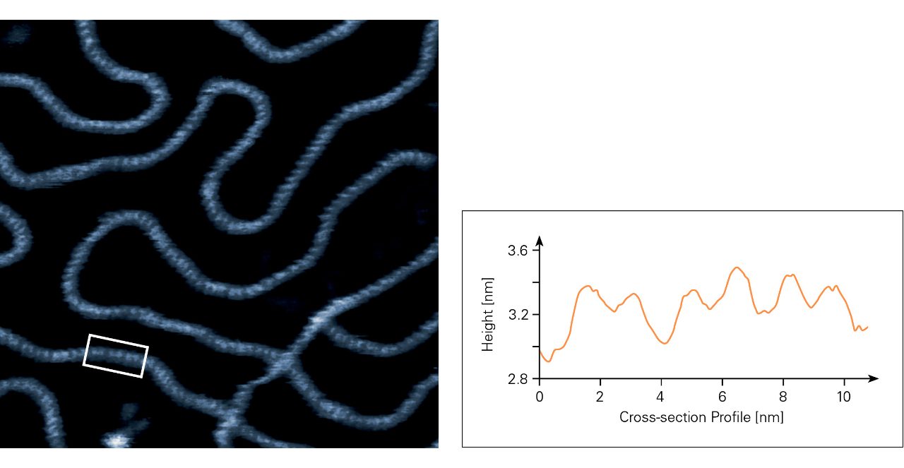
One of the most common applications of AFM is the imaging of DNA. With a diameter of approximately 2 nm, AFM is ideal for providing nanoscale spatial resolution. This enables the visualisation of individual DNA helices and facilitates the study of secondary structures, including torsional variations, bending, and supercoiling. Thus, BioAFM can be used for in-depth analyses of DNA-protein interactions, chromatin organisation, and the mechanical properties of DNA under various physiological conditions. Additionally, BioAFM can operate in liquid environments, making it ideal for observing DNA behaviour in conditions that closely mimic the intracellular milieu [3].
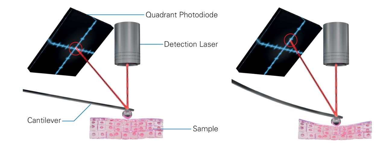
BioAFM Application 2: Fibroblast Rigidity in Clinical Diagnostics
BioAFM can be used in clinical diagnostics to assess cellular rigidity, which is a biomarker in various disease states. For example, fibroblasts exhibit changes in stiffness in response to inflammatory signals, and these changes can correlate with pathological conditions, such as fibrosis or cancer progression. By quantifying the mechanical properties of individual cells, BioAFM enables researchers to explore how fibroblast rigidity relates to disease severity or therapeutic response [4].
BioAFM Application 3: High-Speed BioAFM for Unbiased Measurements
In our podcast with Prof Miles, he also shares that using high-speed BioAFM has the unique advantage of enabling the scanning of larger sample areas with high resolution (or many smaller regions of interest), which means that instead of looking at random small regions, scientists can generate a large image with significantly more data. This means that more data with context and insights can be generated in a fashion that is not biased by region selection.

The Future of AFM
As we look to the future, Prof Miles is excited about 3D data, and the integration of AFM with other imaging techniques. Besides these advances in data acquisition, he also mentioned that automating acquisition using AI could make AFM more widely applicable and less biased.
With companies like Bruker at the forefront of AFM technology and our commitment to innovation, we contribute to AFM development that is impactful and relevant for scientific research for years to come.
You can also download our ressource collection to learn more about AFM technologies and their application.
References
- N. Gavara, “A beginner’s guide to atomic force microscopy probing for cell mechanics,” Microscopy Research and Technique, vol. 80, no. 1, pp. 75–84, 2017, doi: 10.1002/jemt.22776.
- L. Kreplak, “Introduction to Atomic Force Microscopy (AFM) in Biology,” Current Protocols in Protein Science, vol. 85, no. 1, p. 17.7.1-17.7.21, 2016, doi: 10.1002/cpps.14.
- K. H. S. Main, J. I. Provan, P. J. Haynes, G. Wells, J. A. Hartley, and A. L. B. Pyne, “Atomic force microscopy—A tool for structural and translational DNA research,” APL Bioengineering, vol. 5, no. 3, p. 031504, Jul. 2021, doi: 10.1063/5.0054294.
- A. Stylianou, M. Lekka, and T. Stylianopoulos, “AFM assessing of nanomechanical fingerprints for cancer early diagnosis and classification: from single cell to tissue level,” Nanoscale, vol. 10, no. 45, pp. 20930–20945, Nov. 2018, doi: 10.1039/C8NR06146G.
Interested in learning more about this topic?
You can find detailed applications and technical notes, expert-led webinars, and on-demand instrument and measurement demonstrations in our online resource library. Get instant, full-length access to all resources related to this podcast using the form below.
This resource collection includes:
- 1 full-length e-book
- 2 full-length on-demand webinar recordings
- 2 real-time instrument demonstrations