Tech Note: Automating Photomanipulation on the Acquifer IM
Automate large-scale photomanipulation experiments with the
Acquifer IM
In this technical note, readers will learn about the Acquifer IM and photomanipulation module for experiments including photodamaging cells and tissues, switching convertible fluorophores, and performing optogenetic activation. These technologies overcome typical difficulties of photomanipulation by offering an automated approach and simplified workflow control for larger-scale experiments.
Readers can expect to learn more about:
- The photomanipulation module and Acquifer IM provide enhanced scalability for complex photomanipulation experiments
- An optomechnical design enables accurate targeting and manipulation of biological samples
- The Plate-Viewer software on the Acquifer IM high-throughput screening microscope simplifies workflow control
- These technologies support diverse investigations in regeneration and immunology
KEYWORDS: Acquifer Imaging Machine (IM), High-Content Screening, Automated Photomanipulation, Plate-Viewer Software, Cell Biology
In microscopy, photomanipulation refers to using light to control and modify biological processes within cells or tissues. By employing precise wavelengths, intensities, and exposure durations, biological events can be precisely controlled, enabling a range of experimental techniques that provide deep insights into cellular function and dynamics.
Photomanipulation Applications
Photomanipulation is a useful approach due to its spatiotemporal precision, minimal invasiveness to surrounding tissues, and application versatility. However, current photomanipulation technologies are often difficult to implement and are typically restricted to photomanipulation in a few handpicked samples. This impacts the more widespread application of photomanipulation, especially in large-scale assays such as whole organism screening scenarios. This technical note discusses how Bruker's Acquifer Imaging Machine (IM), equipped with the photomanipulation module, provides an ideal solution for automating photomanipulation in high-content screening experiments.
Potential photomanipulation applications include:
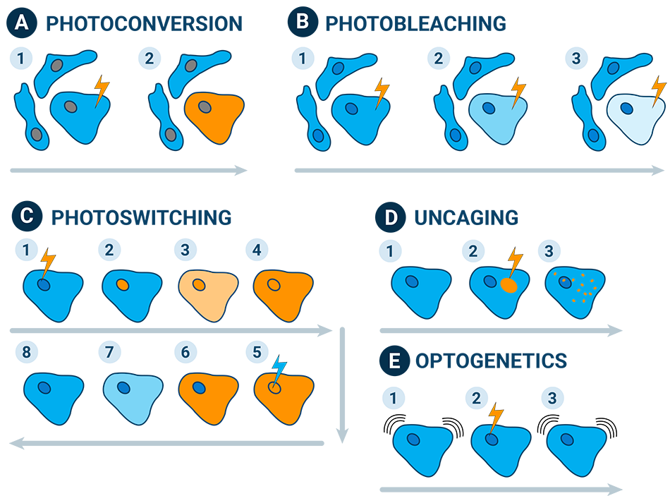
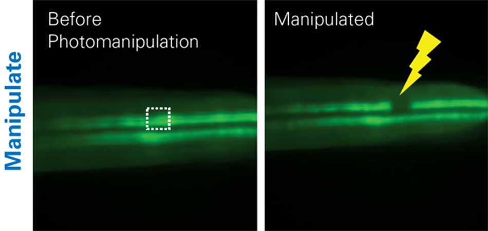
- Photodamage: This involves deliberately using light to damage specific cellular components, such as introducing localized photobleaching to study subcellular dynamics (e.g., fluorescence recovery after photobleaching, also called FRAP1) or introducing largescale tissue damage to study immune response.
- Photoconversion: This uses light to alter the fluorescence properties of certain proteins or dyes from one color to another, such as photoactivatable fluorescent proteins (PA-FPs) or photoconvertible proteins (e.g., Kaede from green to red).
- Uncaging: This uses light to release (or "uncage") bioactive molecules that are initially inactive due to being bound to a "caging" group. Upon exposure to specific wavelengths of light, the caging group is removed, and the molecule becomes active. This can, for example, be applied to spatiotemporally control the uncaging of neurotransmitters or drugs, such as in photopharmacology.2
- Optogenetics: This field combines genetics and optics to control cell activity using light. Introducing light-sensitive proteins (opsins) into specific cells can activate or inhibit cells with light. This can be used to study neural circuits, behavior, cellular processes, and biomaterials with high precision.3
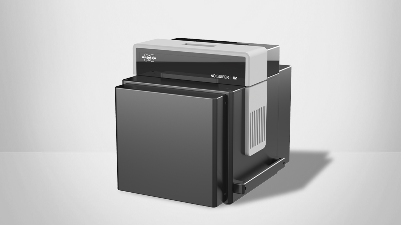
The Acquifer IM Photomanipulation Module
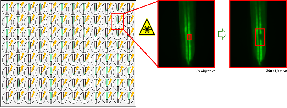
The photomanipulation module is an optional hardware upgrade for Acquifer IM. The Acquifer IM has high-content screening abilities that allow users to easily scale up photomanipulation experiments, including photodamaging cells and tissues, switching convertible fluorophores, uncaging compounds, or performing optogenetic activation in a cell- or tissue-specific manner.
The technology relies on the Acquifer IM's unique optomechanical design with moving optics and a stationary sample holder. This ensures that samples are stationary and that sample movement is minimized, which is particularly important for fragile samples and allows for precise control and imaging results. The incubation chamber can also be cooled down to optimize sample synchronization and reduce sample development, which can lead to unwanted sample movement. This also contributes to lowering sample variability and allows for more consistent results.
The photomanipulation module is automatically positioned under the sample before scanning optics are used for precise photomanipulation. It was developed in collaboration with Rapp Optoelectronic (Wedel, Germany).
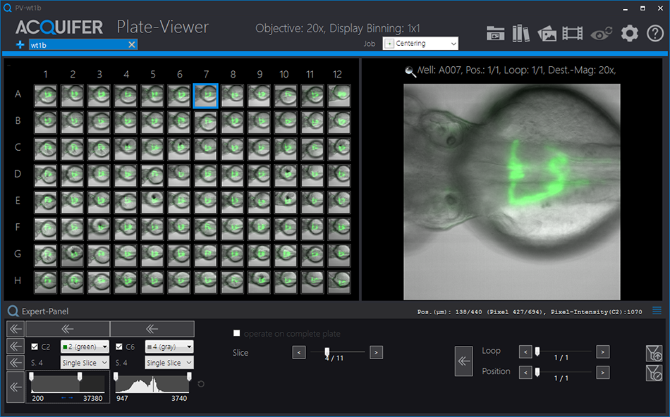
Automating Workflows with the Acquifer Plate-Viewer Enables Easy Scalability
The proprietary Bruker Acquifer Plate-Viewer software simplifies workflow control by integrating smart imaging and supervised feedback microscopy. It allows multiple samples and positions within samples to be annotated and manipulated. In addition, the regions of interest (ROIs) for photomanipulation can take different forms, including lines or polygons, which can be helpful for manipulating within the target shape (such as organs).
A whole dataset can be annotated to set up workflows. The subsequent photomanipulation is then executed automatically, and no further user interaction is required. This is particularly useful when conducting large-scale photomanipulation experiments for biomedical assays, such as drug screens.
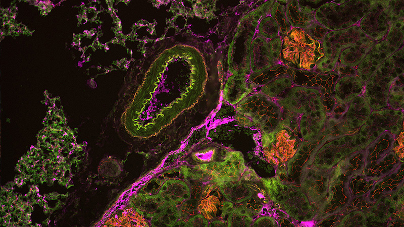
Application Example: Photodamage to Study Kidney Regeneration
Using photomanipulation to induce spatiotemporally controlled tissue damage is ideal for studying regeneration. In the context of kidney regeneration, photodamage can be applied to specific nephron segments or other renal structures, allowing researchers to observe the immediate and long-term cellular responses to injury. This approach facilitates investigating key regenerative processes, such as cell proliferation, differentiation, and the reactivation of developmental pathways.
Moreover, the ability to target damage in a highly controlled manner allows for examining how different cell types within the kidney contribute to the regeneration process. This technique provides critical insights into renal repair's molecular and cellular mechanisms, with potential implications for developing regenerative therapies for kidney diseases.
Workflow Example: Semi-Automated Photodamage to Study Tissue Regeneration
Laser-induced photodamage is a powerful tool for studying tissue regeneration due to its ability to create precise, controlled injuries. This approach allows researchers to induce specific damage in tissues, such as skin, muscle, or organ structures, while precisely controlling the timing and location of the injury. This control is crucial for investigating the dynamic processes involved in tissue repair and regeneration. For instance, in kidney regeneration studies, photodamage can be used to selectively damage the kidney, enabling researchers to observe the cascade of cellular and molecular events that follow.
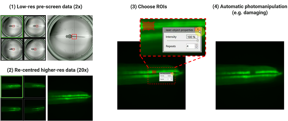
The Acquifer IM can semi-automate high-throughput analysis, which is ideal for:
- Using standardized embryo position with the orientation tool
- Setting up regions of interest with the Plate-Viewer software
- Performing automated photodamaging in user-defined ROIs.
Developing such workflows will be ideal if one wants to set up timelapse imaging overnight and then follow ablation responses and regeneration over time.
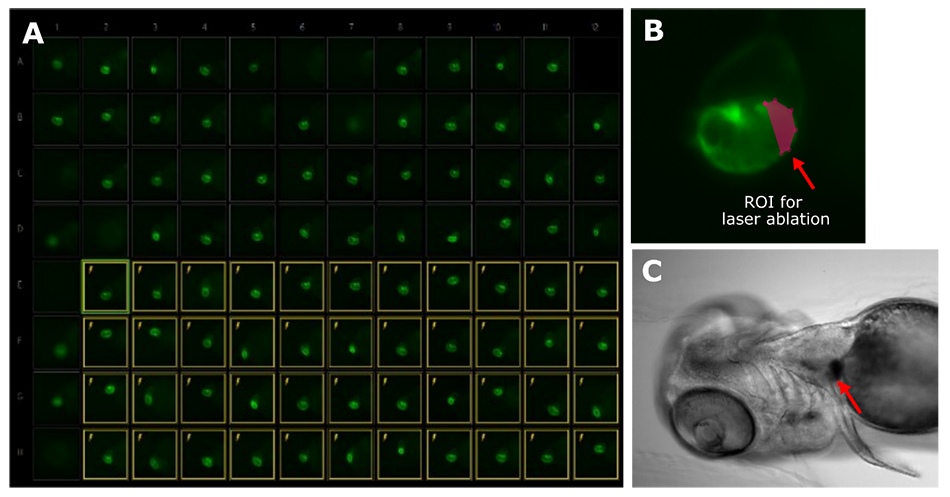
Application Example: Photodamage to Study Fin Immune Response
Inducing photodamage with lasers is a powerful approach for studying immune responses by creating controlled injuries, such as in transparent zebrafish embryos. Photomanipulation allows precise targeting of tissues (developing fins) and observing the real-time behavior of immune cells (tracking the migration of neutrophils to the injury site after the injury).4
This research is crucial as it helps to understand the fundamental mechanisms of immune responses, such as immune cell recruitment and signaling pathways. This can then inform the development of treatments for inflammatory diseases and improve wound healing therapies.
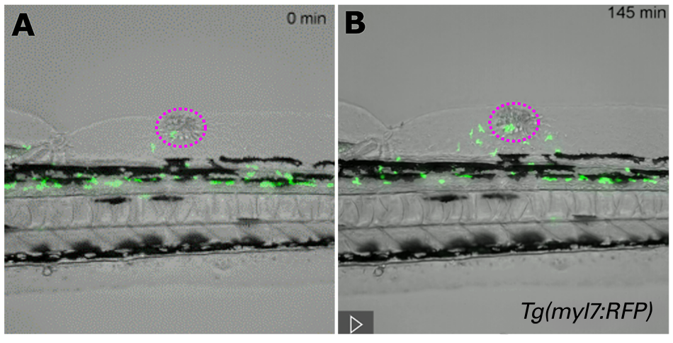
Application Example: Photodamage to Understand Heart Development and Regeneration
Dr. Nadia Mercader’s research focuses on cardiac regeneration, utilizing zebrafish as a model due to their ability to regenerate heart tissue. Her team employs cutting-edge techniques such as laser ablation to target and study specific heart cells, particularly examining the role of epicardial cells in cardiac repair and regeneration. By understanding the interactions between these cells during the regenerative process, Dr. Mercader’s work aims to uncover molecular pathways that could be harnessed to stimulate heart repair in mammals, contributing valuable insights toward developing therapies for heart disease and injury.
Application Example: F-Techniques to Study Subcellular Dynamics
In most biomedical experiments, the fluorescence signal is often solely observed to monitor molecular dynamics, interactions, and cellular processes in real time. However, fluorescence can also be intentionally manipulated to extract more detailed information about subcellular processes.
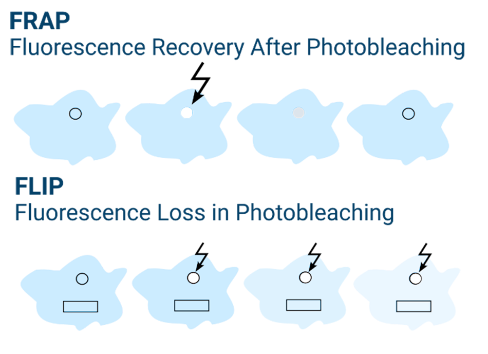
Fluorescence Recovery After Photobleaching (FRAP) is a technique that involves selectively bleaching a specific region of fluorophores within a cell and then monitoring the rate at which fluorescence recovers in that area. By analyzing the recovery dynamics, researchers can gain valuable insights into molecular binding interactions, diffusion rates, and transport mechanisms within the cell.
On the other hand, Fluorescence Loss in Photobleaching (FLIP) examines how the loss of fluorescence propagates throughout the cell following repeated or continuous photobleaching in a specific region. This technique is particularly useful for studying the movement and distribution of molecules within cells, as well as understanding the connectivity between different cellular compartments. Both FRAP and FLIP provide complementary approaches for exploring cellular dynamics and molecular interactions in live cells1.
Solutions for Automated Photomanipulation
Acquifer IM, equipped with the optional add-on photomanipulation module, enables researchers to perform and automate photomanipulation experiments more easily. Acquifer Plate-Viewer software also provides different tools and matching algorithms for selecting ROIs in every well, delivering robust localization of target structures. These innovative technologies open the door for advanced cell biology, biomedicine studies, and more.
References
- H. C. Ishikawa-Ankerhold, R. Ankerhold, and G. P. C. Drummen, “Advanced Fluorescence Microscopy Techniques—FRAP, FLIP, FLAP, FRET and FLIM,” Molecules, vol. 17, no. 4, pp. 4047–4132, Apr. 2012, doi: 10.3390/molecules17044047.
- A. M. J. Gomila and P. Gorostiza, “In Vivo Applications of Photoswitchable Bioactive Compounds,” in Molecular Photoswitches, John Wiley & Sons, Ltd, 2022, pp. 811–842. doi: 10.1002/9783527827626.ch34.
- S. Pearson, J. Feng, and A. del Campo, “Lighting the Path: Light Delivery Strategies to Activate Photoresponsive Biomaterials In Vivo,” Advanced Functional Materials, vol. 31, no. 50, p. 2105989, 2021, doi: 10.1002/adfm.202105989.
- P. Siregar et al., “Optimization of Laser-Based Method to Conduct Skin Ablation in Zebrafish and Development of Deep Learning-Based Method for Skin Wound-Size Measurement,” Inventions, vol. 9, no. 2, Art. no. 2, Apr. 2024, doi: 10.3390/inventions9020025.
Acknowledgements
The Acquifer IM photomanipulation module was developed as part of a cooperation project supported by BMWi’s ZIM program (Central Innovation Programme for SMEs). Partners: Acquifer Imaging GmbH (Heidelberg)/DITABIS AG (Pforzheim), Rapp Optoelectronic (Wedel), Jens Westhoff lab (University Children’s Hospital, Heidelberg), and Holger Erfle lab (IPMB, Heidelberg).
Authors
- Dr Elisabeth Kugler, External Marcom Specialist (E.kugler-extern@bruker.com)
- Dr. Jochen Gehrig, Senior Product and Applications Specialist (Jochen.gehrig@bruker.com)
- Melissa Martin, Life Science Writer (Melissa.martin@bruker.com)
©2025 Bruker Corporation. Acquifer IM and Plate-Viewer are trademarks of Bruker. All other trademarks are the property of their respective companies. All rights reserved. TN2603, Rev. B0.