Destriping Module
Due to the orthogonal illumination-detection geometry, light-sheet microscopes are prone to artifacts such as shadows and stripes. These artifacts are caused by the illumination laser being absorbed or scattered by inhomogeneities in the sample, such as cell nuclei or pigments. Due to this light interaction with matter, shadows are cast behind the object, causing stripe artifacts.
In an ideal scenario, 100% of the illumination light would transmit through the sample of interest. However, light interacts with matter and impacts transmission by absorption, diffraction, reflection, refraction, and scattering. Shadow effects can lead to stripe artifacts, which can be reduced by pivoting the illumination light sheet.
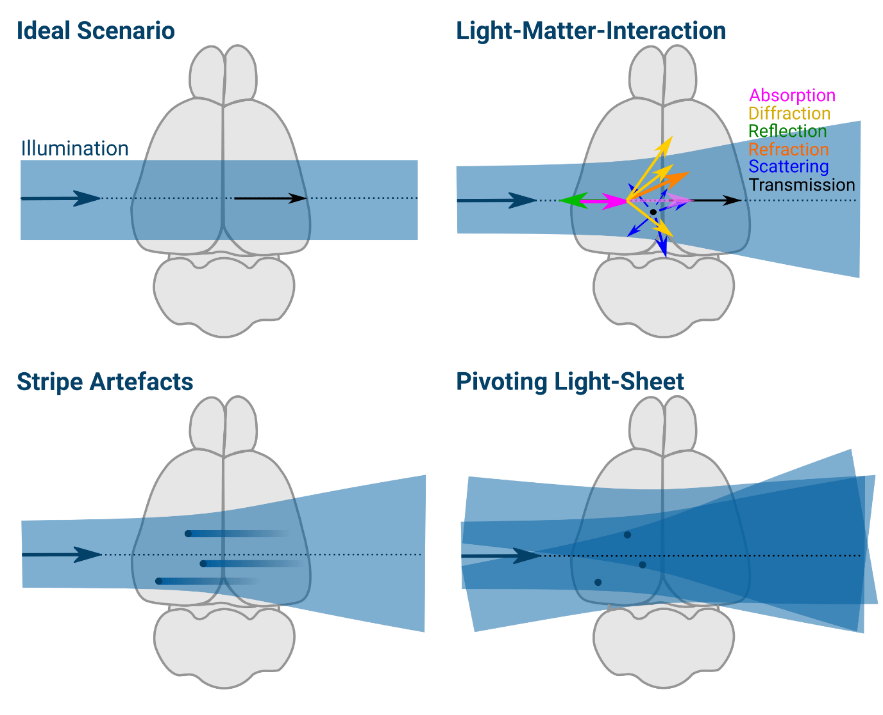
▲ In an ideal scenario, light would penetrate 100% of a sample, but light-matter interaction, including absorption, diffraction, reflection, refraction, scattering, and transmission interfere with light transmission.
How to avoid and remove stripe artifacts?
Several approaches can help reduce stripe artifacts, including:
- Genetic or chemical inhibition of tissue formation (e.g., Nacre zebrafish or PTU treatment)
- Depigmentation, or tissue bleaching
- Tissue clearing
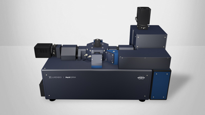
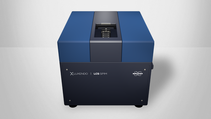
- Multiple illumination sources: Opposing light sources and multi-view acquisition are supported by Bruker's LCS SPIM, TruLive3D Imager, and MuVi SPIM light-sheet microscopes
- Pivoting the illumination light-sheet
- Algorithmic destriping
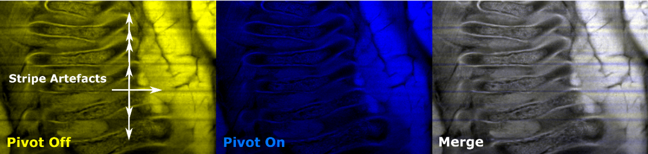
▲ Cleared mouse embryo. Left: Image collected without the destriping module showing striping artifacts. Middle: Image collected with the destriping module showing successful removal of striping artifacts. Right: Overlap shows blue stripes, i.e., the missed regions without pivoting.
How is Destriping achieved with Bruker Light-Sheet Microscopes?
Bruker's light-sheet microscopes achieve destriping by pivoting the illumination light-sheet in the focal plane of the detection objective around its beam waist, allowing the illumination beam to reach the same point of the tissue under different angles as it oscillates. As the oscillation is much faster than the SPIM camera exposure time, a homogenous illumination is generated, thus minimizing potential striping artifacts without compromising acquisition speed.
The destriping module is an add-on for the MuVi SPIM and TruLive3D Imager and is built-in on the LCS SPIM.