TruLive3D Imager

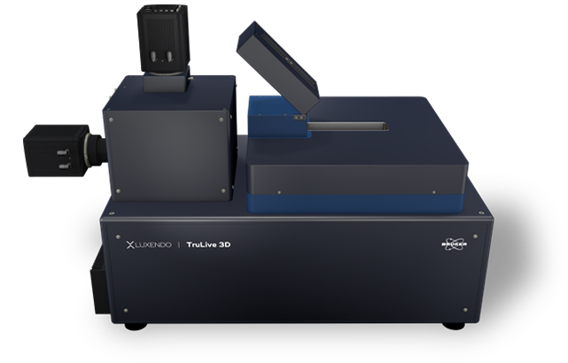
TruLive3D Imager
The Bruker Luxendo TruLive3D Imager is optimized for fast 3D multi-sample volume imaging of delicate live specimens in their native 3D environment. Thus, it is particularly well-suited for time-lapse and high-throughput imaging of 3D spheroids, organoids, 3D cell cultures, and small embryos. Using an optical concept, with dual-sided illumination and single-lens detection from below, enables fast acquisition speed, high-resolution imaging, and minimal shadowing effects.
Latest technology advances allow for state-of-the-art uniform illumination and the optional addition of a photomanipulation module.
Compact and Vibration-Free Design for Long-Term In Vivo Experiments
The TruLive3D Imager has a compact, robust and vibration-free design that provides maximal stability even during long-term and high-throughput experiments. Like all Bruker Luxendo's light-sheet solutions, the TruLive3D Imager is tailored to fit onto a lab bench, without the need for an air table. The proprietary piezo-crawler stages ensure longevity and precision for permanently accurate specimen positioning.
As the TruLive3D Imager is optimized for fast, long-term in vivo imaging, the system has advanced environmental control. This allows unparalleled imaging of experiments such as:
- Spheroid and organoid cell-divisions, cell-interactions, and dynamics
- 3D cell culture growth and drug responses
- Small early embryo development, such as mouse, zebrafish, and drosophila
Dual-Destriping: Impressive Elimination of Striping Artifacts

When imaging, obstacles like pigments or cell nuclei can interfere with light, leading to images with shadows and striping artifacts. The next-generation SPIM employs dual-destriping via pivot scanning, which generates a homogeneous illumination profile without compromising acquisition speed.
TAG-Lens: Light-Sheet Length Control with Fast Axial Scanning
Thanks to state-of-the-art technologies, ultra-fast varifocal lenses can be used to fine-tune the system's optics. Luxendo SPIMs use fluid-filled devices that are driven by an acoustic wave, called tunable acoustic gradient lenses (or TAG lenses), for adaptive optics. These TAG lenses allow better light-sheet control and uniformity while reducing axial stretching.
Deep and Crisp Imaging

Illumination and Detection
Using an inverted setup with dual-side illumination, single-lens detection, and dual-destriping, the TruLive3D Imager allows for minimal shadowing effects, uniform illumination, and deep sample penetration. In addition to advanced geometry, the TruLive3D Imager can achieve a high resolution.
While low phototoxicity is a key characteristic of light-sheet microscopy in general, the TruLive3D Imager takes sample-preserving imaging to the next step.
Native and Natural
An important goal in microscopy is sample health and integrity. To support this, the TruLive3D Imager is equipped with environmental control that supports long-term imaging (hours to days). In addition to a generous sample chamber for immersion medium, this includes control over:
- Temperature 20 to 37°C
- Gas concentration (CO2, O2, N2)
- Humidity

Multiplexing

Large Sample Holder
To take organoid, spheroid, or 3D cell culture imaging to the next level, the TruLive3D Imager fits a large sample holder (length = 75 mm), which can accomodate hundreds of samples. Additionally, the new TruLive3D Dish series allows the user to separate the sample holder to student different experimental conditions at once. This setup makes the TruLive3D Imager ideal to study multiple samples over long periods of time and under different experimental conditions. An additional wide-field imaging option facilitates fast sample positioning.
Sample Embedding
Easy and intuitive sample embedding is key for enabling scientists to excel in their research. While the large sample holder (length = 75 mm) can accommodate hundreds of samples, the TruLive3D Dish series seamlessly integrates into the system and allows for testing of different conditions. Each TruLive3D Dish contains two sample spaces and three disposable dishes that extend the capacities of the system. Thus, a total of six experimental setups can be tested at once. For sample embedding, all holders use Fluorinated ethylene propylene (FEP) foil, which functions as a physical barrier and a "curved coverglass."
The FEP foil lining the holder serves dual functions as a “curved coverglass” and as a physical barrier between the immersion medium and sample medium. Samples can be raised directly on the foil or transferred, e.g., with a drop of matrigel. Importantly, only small volumes are needed for drug and compound testing, making the system highly effective. Using two LEDs and an overview camera allows easy sample localization and experiment setup through an overview camera looking from the side.
TruLive 3D Imager Design


Advanced Multiplex Acquisition
With the ability to study hundreds of samples at once and to perform long-term imaging for developmental and tissue studies, the TruLive 3D Imager provides a unique multiplex setup. To improve efficiency, we offer the option to partition the sample chamber, allowing researchers to study multiple drug treatments in parallel. This approach can save valuable time and resources, as time is often the scarcest commodity in research.
Innovative and Easy Sample Handling
The design of the TruLive 3D Imager allows for easy access from above and allows for swift immersion medium changes, which supports long-term imaging. Additionally, the system comes equipped with two LED illuminations which can be used for both sample positioning and experimental setup. These features can help streamline the imaging process and improve overall research productivity.
Long-Term Imaging
The TruLive 3D Imager is a state-of-the-art imaging system specifically designed for long-term imaging purposes. With its advanced features, the TruLive 3D Imager can acquire data over extended periods of hours and days all while ensuring the preservation of sample health and integrity. Its high quantum efficiencies of up to 82% provide excellent image quality, and its stage travel ranges of 66 x 3 x 3 mm allow for precise sample positioning. Additionally, this imager offers a wide range of final magnification options from 8x to 62.5x, making it a versatile tool for various research applications.
Organoids and Spheroids
Diabetes is a global health burden so understanding pancreas development and health is critical to learn more about the disease and treatment options. Using pancreatic spheres, one can study how cell fate choices and organ architecture play a role in pancreas health and disease.
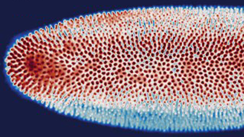
Pancreatic Sphere
hESC-derived pancreatic spheres. Imaging on the TruLive3D Imager enables collecting information of several samples in one experiment. Visualization: Imaris (Bitplane)
Courtesy of:
Yung Hae Kim
Grapin-Botton Group, MPI-CBG
Dresden, Germany
Photomanipulation for Cell and Tissue Biology
Understanding how the immune system works and the various processes of immune defense is critical to understand health, disease, and vaccine efficacy. Often, immune system dynamics are impossible to measure with conventional approaches and need to be studied in vivo. This makes it a unique challenge for light-sheet microscopy.

Timelapse of cytokine dynamics in a tail wound assay. Pseudo-Brightfield and il1b:GFP (green) merged. Captured every 5 min for 12 hours.
Sample courtesy of: Dr. Elizabeth Jerison, Stanford University.
Tumorigenesis
Tumors are known to establish their own tissue ecosystem habitats. However, how tumors arise from a few malignant cells, how they interact with surrounding cells, and what this means for cancer progression is unknown. Having the ability to study tumorigenesis over time in a controlled system provides new insights into the decisions and processes of tumorigenesis.
Cell Biology in a Dish
The neurovascular unit is a critical component of the nervous system. However, the role of components such as glial cells is still not fully understood. Particularly brain glial cells, called astrocytes, play many different roles in neuron health and function. Thus so, understanding neuro-glial interactions is critical to understanding nervous system health. Reducing this system to "cell biology on a dish" adds control over the system, while enabling toxicological studies.
Specifications
| Objective | Tube Lens | Magnification | Pixel Size [nm] | FOV [µm] |
|---|---|---|---|---|
| Nikon CFI APO LWD 25x 1.10 NA | 100 | 12.5x | 520 | 1065 |
| 200 | 25x | 260 | 532 | |
| 400 | 50x | 130 | 266 | |
| Olympus XLUMPLFLN 16x 0.8 NA | 100 | 8x | 813 | 1664 |
| 200 | 16x | 406 | 832 | |
| 400 | 32x | 203 | 416 | |
| 500 | 40x | 163 | 333 |
Illumination Optics
- Chromatic correction from 440 to 660 nm
- Light-sheet generation by beam scanning
- Flexible light-sheet thickness (2 µm to 6 µm)
- Motorised ND filter set (90%, 10%, 1%) for high flexibility in using low laser power
- Two Nikon CFI Plan Fluor 10x W 0.3 NA water immersion objective lens
Detection Optics
- One Nikon CFI Apo 25x W 1.1 NA water immersion objective lens
- Two spectral detection channels, each equipped with a fast filter wheel (10 positions and 50 ms switching time between adjacent positions) Filters adapted to the selected laser lines
- Three motorised dichroic mirror positions for simultaneous dual channel acquisition
- Motorised collar ring controlTwo high-speed sCMOS cameras Hamamatsu Orca Flash 4.0 V3
- Maximum frame rate >80 fps at full frame (2048 × 2048 pixels of 6.5 µm × 6.5 µm size) and up to 500 fps at subframe cropping
- Peak quantum efficiency (QE): 82% @ 560 nm
System Comparison
* COMPATIBLE MODULE
| The perfect allrounder - multi-view imaging for diverse samples (live and cleared option) | Multi-sample and dual view flexibility for highly sensitive imaging, ideal for organoids and 3D cell cultures | In vivo multi-sample and multi-condition imaging, ideal for drug treatment comparison | Perfection for cleared samples prepared by any type of clearing protocol | High-end imaging with various beam patterns from single cells to 3D cell cultures | |
|---|---|---|---|---|---|
| Geometry | Horizontal with multiview | Horizontal with dual view | Inverted with dual illumination | Inverted dual illumination with moving optics | Inverted geometry at 360° |
| Beam Type | Gaussian | Gaussian and Bessel | Gaussian | Gaussian | Advanced Illumination Module (AIM) |
| Application Examples | - Long-term imaging of Drosophila development - Photomanipulation studies in zebrafish embryos - Cleared and stained samples up to mouse brain size | - High-resolution full 3D imaging of multiple organoids over time - Long-term imaging of highly light-sensitive samples - Comparing the pharmacological impact in zebrafish wound healing after PM | - Time-lapse imaging of multiple pancreatic spheres - Studying pharmacological impact on zebrafish development - Comparing genetically altered organoids | - Pharmacokinetics in mouse brain - Multi-stained rat brain - Whole mouse imaging | - Fast imaging of photo-sensitive 2D cell culture - Lattice imaging for enhanced resolution in 3D cell cultures - FCS and FLIM in cell culture |
| User Level | Beginner friendly | Beginner friendly | Beginner friendly | Intermediate | Advanced |
| # Lenses | 4 lenses (2 IO, 2 DO) | 3 lenses (1 IO, 2 DO) | 3 lenses (2 IO, 1 DO) | 3 lenses (2 IO, 1 DO) | 2 lenses (1 IO, 1 DO) |
| Multi-View | ✓ | ✓ | |||
| Live/Fixed Samples | ✓ | ✓ | ✓ | ✓ | |
| Cleared Samples | ✓ | ✓ | |||
| Expanded Samples | ✓ | ✓ | ✓ | ✓ | ✓ |
| Best Embedding | Capillary/FEP tube w/ agarose 3D stage | Custom-solution and FEP foil | TruLive3D dishes and FEP foil | Quartz-crystal cuvette | FEP foil; glass slides |
| Photomanipulation* | ✓ | ✓ | ✓ | ✓ | |
| Environmental Control* | ✓ | ✓ | ✓ | ✓ | |
| Destriping/Uniform Illumination* | ✓ | ✓ | ✓ | ✓ | ✓ (w/ Advanced Illumination Module) |
| Benchtop Design | ✓ | ✓ | ✓ | ✓ | ✓ |
Accesories

TruLive3D Dishes

TrueLive3D Imager Sample Holder
Software

Luxendo's intuitive user interface offers a simple setup and execution of multidimensional experiments, while real-time control is handled by an embedded controller to ensure microsecond-precision timing independent of the PC’s performance fluctuations.
Precise timing control of all connected devices is a prerequisite for reliable experimental outcomes. Full control of data streaming to storage as well as GPU-supported image processing further complements the overall performance.
Electronics, Microscope Software and Computer
- Embedded microscope software with an open communication interface: documented API, TCP/IP-based communication
- Flexible GUI for interactive microscope control and experiment design
- Computer with 256 GB RAM, Intel dual 8 core CPU
- High-speed RAID controller for data streaming, 8×4 TB local storage in RAID 0
- GeForce RTX 3070 graphics card
- 10 Gbit/s on-board Ethernet port
- 1 UHTV 43 inch display
All-in-One LuxBundle Software
Intuitive design
Bruker's LuxBundle software saves time and enhances productivity by providing:
- All-in-one, easy-to-use interface for acquisition, viewing, and post-processing
- Fully scriptable microscope control and post-processing via open interface (e.g., Python or any other language), ready for custom "smart" microscopy
- High reproducibility of experiments: all parameters are saved in the metadata and configurations can be saved for future experiments
- Data formats (.tiff, .hdf5, .ims) compatible with common image processing software: Imaris, Aivia, BigDataViewer, Arivis, Fiji, Python, Matlab, Napari
Impressive Image Post Processor
Our dual-view light-sheet microscopes record a sample from different angles/views and generate images composed of multiple tiles. The LuxBundle software ensures high-quality, 360° crisp images of the sample that compensate for absorption and scattering. Features include:
- Multi-color alignment
- Tile stitching of hundreds of tiles for large samples
- Multi-view image fusion and deconvolution
3D Data Viewer
LuxBundle's integrated 3D data viewer allows researchers to inspect the entire dataset directly after acquisition. This gives users control over their data with key capabilities, including:
- The ability to turn tile stitching on or off
- Both raw and post-processed images
- Fast viewing of multi-terabyte data sets
- Flexible options to draw and annotate regions and landmarks